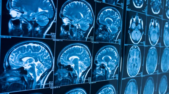
New technique pushes closer to real-time MRI brain imaging
A team of researchers backed by a division within the National Institutes of Health (NIH) has created a novel MRI technique that can image a thinking brain 60-times faster than traditional methods.
The imaging method measures changes in the brain’s tissue stiffness, tracking brain function on a time scale of 100 milliseconds, according to a statement from the National Institute of Biomedical Imaging and Bioengineering (NIBIB), which provided the study grant. Traditionally, researchers use fMRI to measure brain activity, but mounds of important information are lost due to long brain-stimulus response times.
“This study has the potential to revolutionize the way scientists study brain diseases,” said Krishna Kandarpa, MD, PhD, director of research science and strategic directions at NIBIB. “Developing a new MRI technique relies heavily on physics and engineering principles, which are areas in which NIBIB investigators excel. The results would have been hard to achieve without the collaboration of this team of experts.”
For the study, the initial plan was to use magnetic resonance elastography (MRE)—which quantifies tissue stiffness by measuring the speed at which mechanical vibrations move through tissue—along with another MRI method to study scar tissue in patient’s lungs.
After encountering a few complications, the team decided to start with a mouse brain. And while experimenting with a few different methods, the researchers settled on one that switches between “on and off stimulus states” created by an electrical impulse sent to the mouse’s rear limb every nine seconds. Two brain images are generated, one from each stimulus state, which are then subtracted to reveal changes in tissue stiffness.
The positive results prompted the team to try faster speeds, ultimately determining that 10% changes in tissue stiffness were apparent every 100 milliseconds.
Similar studies have already been performed in healthy human brains; preliminary results show changes in tissue stiffness in as little as 24 milliseconds. This, the statement read, is the closest to real-time MRI brain imaging researchers have achieved to date.
The full study can be read in Science Advances.

