UK researchers use holograms to train future interventional radiologists
There is a growing need for new approaches to train medical students, and a novel method utilizing holograms may be just what radiologists are looking for.
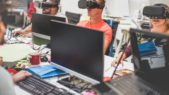
A group of experts from Imperial College London’s Faculty of Medicine and Digital Learning Hub is using Microsoft HoloLens2 to teach interventional radiology trainees how to perform complex procedures, promising improved technical skills in a realistic environment. The work builds on the mixed-reality approach that has previously helped surgeons peer through virtual tissue to perform reconstructive surgery.
“By building the simulation in mixed-reality it not only allows for accurate imaging placement, but also for crucial tactile feedback,” Mohamad Hamady, MD, professor at Imperial College, said in a news item.
During the training course, students practice simulations of image-guided needle biopsies. The virtual environment is built from real-world patient images and allows wearers to use real needles and scan as if they were performing an actual CT-guided procedure, the authors noted.
Microsoft HoloLens was approved by the FDA in 2018 for use in pre-operative surgical planning. HoloLens2 is the second version of the headset and enables an individual to interact with virtual “holograms.”
The current trial involves junior interventional radiology trainees from various centers across London, according to the college. Researchers believe the approach can shorten the required training time for this particular group of radiologists—a specialty growing in demand, they noted.
"This app really demonstrates the potential of mixed reality to provide high-fidelity experience at the fraction of the cost of traditional methods,” Dimitri Amiras, MD, a musculoskeletal radiologist at the college said. “As technology improves this will not only change the way we train but also the way in which we perform procedures.”

