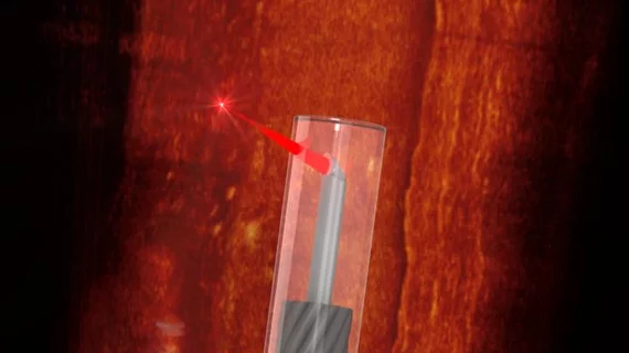
‘World’s smallest’ imaging device has large implications for cardio care
A team of international researchers has used 3D printing to create what they believe is the world’s smallest imaging device, with plans to help treat heart disease.
German and Australian scientists teamed up to create the optical coherence tomography endoscope, sharing their development July 20 in Light: Science & Applications. The device is so small the group was able to scan inside the blood vessels of mice.
Co-author of the study, Jiawen Li, PhD, with the University of Adelaide in Australia, and colleagues now hope to turn their probe toward better understanding cardiovascular disease, which kills one Australian every 19 minutes.
"A major factor in heart disease is the plaques, made up of fats, cholesterol and other substances that build up in the vessel walls," added Li, with the university’s Institute for Photonics and Advanced Sensing. “Miniaturized endoscopes, which act like tiny cameras, allow doctors to see how these plaques form and explore new ways to treat them.”
The team listed off a number of other potential applications for their creation, which has a diameter of 0.46 millimeters. That includes safe access to the internal carotid artery and its branches, where 85% of intracranial aneurysms arise, the authors noted in the study.
New pulmonary applications, including guiding biopsy in peripheral nodules and imaging inside of the cochlea in the ear, are also “now within reach.”
“It's exciting to work on a project where we take these innovations and build them into something so useful,” Li added in a Tuesday statement. “It's amazing what we can do when we put engineers and medical clinicians together.”

