Artificial Intelligence

Artificial intelligence (AI) is becoming a crucial component of healthcare to help augment physicians and make them more efficient. In medical imaging, it is helping radiologists more efficiently manage PACS worklists, enable structured reporting, auto detect injuries and diseases, and to pull in relevant prior exams and patient data. In cardiology, AI is helping automate tasks and measurements on imaging and in reporting systems, guides novice echo users to improve imaging and accuracy, and can risk stratify patients. AI includes deep learning algorithms, machine learning, computer-aided detection (CAD) systems, and convolutional neural networks. 

Thumbnail

4D MRI reliable for diagnosing parathyroid tumors

Four-dimensional (4D) MRI with dynamic contrast-enhanced (DCE) sequencing is a reliable method for localizing parathyroid lesions, reported authors of a single-center study published in the European Journal of Radiology.

Thumbnail

AI saves radiologists time by triaging chest x-rays, but is it clinically feasible?

There’s been plenty of hype around the potential of artificial intelligence (AI) to ease radiology workloads. And a new convolutional neural network approach detailed in a Jan. 22 study greatly reduced reporting backlog by accurately triaging chest x-rays in real-time.

Thumbnail

How imaging, 3D printing gave one man a new face

On Jan. 6, medical imaging and three-dimensional (3D) printing went above and beyond when Eduardo Rodriguez, MD, a plastic surgeon and face transplant specialist at NYU Langone Medical Center in New York City, used the technologies to help him perform his third face transplant surgery, according to a recent report by Popular Science.

Thumbnail

Australian researchers’ ‘ultra’ ultrasound could boost sensitivity of standard imaging

Researchers from the University of Queensland in Australia have developed “ultraprecise ultrasound” sensors that are sensitive enough to hear the formation of surrounding air molecules, according to new research published online Jan. 10 in Nature Communications.

Thumbnail

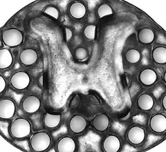
3D-printed implant created from MRIs may help treat spinal cord injuries

Using 3D printing, researchers from the University of California San Diego created spinal cord implants modeled from MRI scans that support nerve cell growth in spinal cord injuries and help restore lost physical mobility, according to a new study published online Jan. 14 in Nature Medicine.

Thumbnail

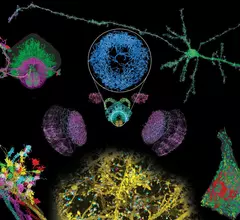
Novel microscopy method enables high-resolution study of neural brain circuits

A novel imaging technique combining two microscopy methods allowed researchers to visualize neural circuits across the brain at four-times that of typical resolutions, according to research published Jan. 17 in Science. The approach can be completed much faster than previously thought possible.

Thumbnail

3D imaging of tissue oxygenation may improve cancer treatments

“Knowing about the oxygenation (of tissues) might allow us to tailor the therapy to make it better and to use other therapeutic interventions that are more appropriate,” said Vikram Kodibagkar, PhD, an associate professor of engineering at Arizona State University in Tempe, according to a recent university news release.

Thumbnail

California team gains insight into how the brain stores time-related information

A team at the University of California, Irvine (UCI) gained new insights into how the brain stores information related to time with the help of fMRI and HBO’s “Curb Your Enthusiasm.”

Around the web

Positron, a New York-based nuclear imaging company, will now provide Upbeat Cardiology Solutions with advanced PET/CT systems and services. 

The nuclear imaging isotope shortage of molybdenum-99 may be over now that the sidelined reactor is restarting. ASNC's president says PET and new SPECT technologies helped cardiac imaging labs better weather the storm.

CMS has more than doubled the CCTA payment rate from $175 to $357.13. The move, expected to have a significant impact on the utilization of cardiac CT, received immediate praise from imaging specialists.