Artificial Intelligence

Artificial intelligence (AI) is becoming a crucial component of healthcare to help augment physicians and make them more efficient. In medical imaging, it is helping radiologists more efficiently manage PACS worklists, enable structured reporting, auto detect injuries and diseases, and to pull in relevant prior exams and patient data. In cardiology, AI is helping automate tasks and measurements on imaging and in reporting systems, guides novice echo users to improve imaging and accuracy, and can risk stratify patients. AI includes deep learning algorithms, machine learning, computer-aided detection (CAD) systems, and convolutional neural networks. 

Thumbnail

CT colonography effectively identifies carpet lesions

CT colonography has been found to effectively depict carpet lesions, according to a study published in the February issue of Radiology.

Thumbnail

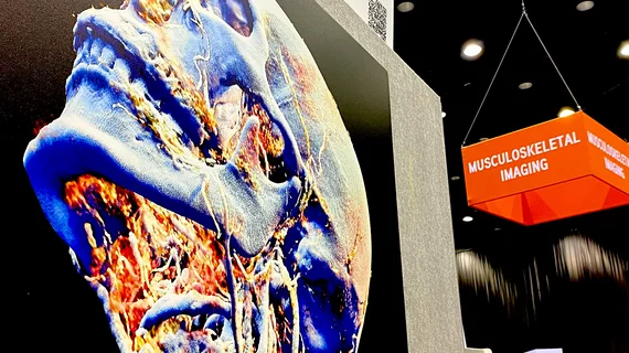
MRI’s Expanding Frontiers: Scanning More, Scanning Faster

Take a look at the latest in PET/MR, parallel imaging and more.

Thumbnail

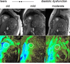
Myocardial relaxation diagnosis possible with SWA for cardiac MR elastography

Low frequency shear-wave amplitude (SWA) vibration for cardiac MR elastography offers valuable diagnostic information on myocardial relaxation, according to a study published online Jan. 19 by Radiology.

Thumbnail

CT attenuation offers insight into pancreatic fat, diabetes

CT attenuation indexes are correlated with pancreatic fat and associated with pancreatic dysfunction that could lead to diabetes, according to a study published online Jan. 16 in Radiology.

Thumbnail

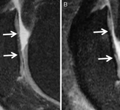
CaLS system reproducible, improves cartilage lesion detection rates

The CaLS system, a reproducible scoring system for cartilage lesions, has been found to improve the detection rate for disease progression monitoring, according to a study published online Jan. 16 by Radiology. 

Thumbnail

Pair of CADx programs show off their stuff

Computer aided detection (CADx) programs offer lots of potential to help radiologists avoid missing important findings, but the benefits vary between applications. Two recent studies, however, have given high marks to CADx programs for identifying pulmonary embolisms (PEs) and breast lesions.

Thumbnail

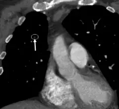
CADx catches missed pulmonary embolisms

A prototype computer aided detection (CADx) program for identifying pulmonary embolisms (PEs) on CT angiography was able to detect a PE in more than 75 percent of cases in which an acute PE had been missed in clinical practice, according to a study published in the January issue of the American Journal of Roentgenology.

Thumbnail

Iterative model reconstruction lowers dose in cardiac CT

Knowledge-based iterative model reconstruction (IMR) can provide improved image quality at super-low-dose cardiac CT with 20 percent of the standard tube current, according to a study published in the January 2014 issue of Academic Radiology. 

Around the web

RBMA President Peter Moffatt discusses some of the biggest obstacles facing the specialty in the new year. 

Deepak Bhatt, MD, director of the Mount Sinai Fuster Heart Hospital and principal investigator of the TRANSFORM trial, explains an emerging technique for cardiac screening: combining coronary CT angiography with artificial intelligence for plaque analysis to create an approach similar to mammography.

A total of 16 cardiology practices from 12 states settled with the DOJ to resolve allegations they overbilled Medicare for imaging agents used to diagnose cardiovascular disease.