Magnetic Resonance Imaging

Cardiac magnetic resonance imaging MRI is used as both a functional and anatomical cardiac imaging test. It offers excellent soft tissue detail and the ability to quantify cardiac function. MRI scans can be performed with or without gadolinium contrast depending on what information is needed. Unlike computed tomography (CT), MRI does not use X-ray radiation, but patients with metal implants may have contraindications for MRI use because MR will heat up most metal objects. MRI exams usually take much longer than CT scans. How does MRI work? MR creates images by using powerful magnets to polarize hydrogen atoms in water (the body is made of of more than 80% water) so they face in one direction. A radiofrequency pulse is then used to ping these atoms, causing them to wobble, or resonate. The MRI coils detect this and computers can assemble images from the signals. Basic MRI scans will focus on the resonance of fat and water in two different sequences, which highlight and contrast different features in the anatomy.
Thumbnail

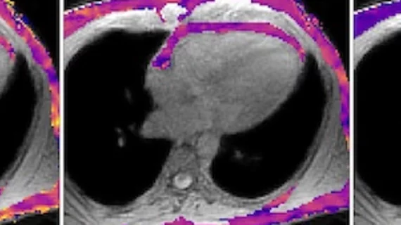
T2 mapping may uncover cardiotoxic marker early enough to prevent heart failure

T2 mapping derived from weekly cardiac MRIs helped researchers identify cardiotoxicity at an early and reversible stage, a finding which may have implications for cancer patients at risk of chemotherapy-induced heart failure.

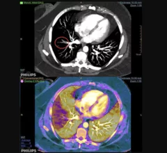
Example of spectral, or dual-energy CT, confirming a pulmonary embolism (PE). Image courtesy of Philips Healthcare

MRA vs CTA for evaluating pulmonary embolism: Does the chosen modality impact downstream imaging utilization?

MR angiography (MRA) is a relatively new alternative to CT angiography (CTA) for the evaluation of suspected pulmonary embolism (PE) and it even has one major advantage over CTA: it does not expose patients to ionizing radiation.

Example of pulmonary embolism (PE) detection on CT and confirmed using spectral CT. Image courtesy of Philips Healthcare

Researchers 'concerned' CT angiography topped MRA in follow-ups for pulmonary embolism

A recent study found whether a patient received MR angiography (MRA) instead of CT angiography (CTA) for a pulmonary embolism (PE) had little effect on the rate of follow-up chest CT or MRI one year later.

Image reconstruction algorithm, MRI-derived heart strain values can aid prognosis in amyloidosis patients

Recent research found strain parameters taken from a cine MRI-based deformable registration algorithm (DRA) can determine the severity of amyloid buildup in the heart and may provide prognostic information on patients with light-chain (AL) amyloidosis.

Thumbnail

Seeing the Big Picture: Training Today’s Imagers to ‘Think Multimodality’

Cardiologists are receiving more exposure to different imaging modalities during their fellowships, but their job prospects and training vary widely. A more comprehensive and multimodality training approach could lead to better results.  

Around the web

GE HealthCare designed the new-look Revolution Vibe CT scanner to help hospitals and health systems embrace CCTA and improve overall efficiency.

Clinicians have been using HeartSee to diagnose and treat coronary artery disease since the technology first debuted back in 2018. These latest updates, set to roll out to existing users, are designed to improve diagnostic performance and user access.

The cardiac technologies clinicians use for CVD evaluations have changed significantly in recent years, according to a new analysis of CMS data. While some modalities are on the rise, others are being utilized much less than ever before.