Magnetic Resonance Imaging

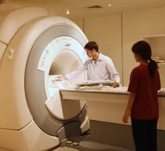
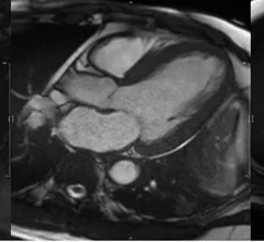
Cardiac magnetic resonance imaging MRI is used as both a functional and anatomical cardiac imaging test. It offers excellent soft tissue detail and the ability to quantify cardiac function. MRI scans can be performed with or without gadolinium contrast depending on what information is needed. Unlike computed tomography (CT), MRI does not use X-ray radiation, but patients with metal implants may have contraindications for MRI use because MR will heat up most metal objects. MRI exams usually take much longer than CT scans. How does MRI work? MR creates images by using powerful magnets to polarize hydrogen atoms in water (the body is made of of more than 80% water) so they face in one direction. A radiofrequency pulse is then used to ping these atoms, causing them to wobble, or resonate. The MRI coils detect this and computers can assemble images from the signals. Basic MRI scans will focus on the resonance of fat and water in two different sequences, which highlight and contrast different features in the anatomy.
Thumbnail

No need for updated imaging prior to major surgery in some cases, new data show

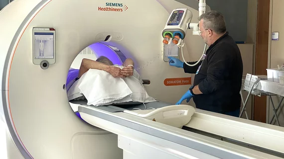
Prior chest imaging can sometimes be just as predictive of adverse events after surgery as updated imaging, according to new data set to be presented at the annual conference of the American College of Cardiology on March 5. 

Thumbnail

Fatal accident calls MRI safety standards into question—are more regulations needed?

There are a number of well-intended recommendations for preventing MRI injuries, but many of them stop short of implementing safety requirements.

mixed reality LAAO Chase Western Reserve MRI

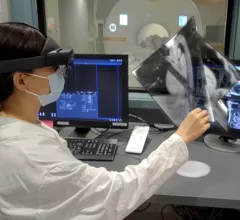
Researchers receive $3.7M to attempt robotic heart surgery inside MRI scanner

The group—which includes engineers, cardiologist, radiologists and other specialists—will attempt to perform a robotic-controlled left atrial appendage occlusion (LAAO) on a patient inside an MRI scanner.

Thumbnail

New cardiac MR technique cuts scan time by more than half

Using a new 4D flow cardiac MRI scan, experts were able to complete cardiac examinations in just eight minutes.

Thumbnail

Cardiac MRI reveals that even competitive athletes are subject to reduced cardiac function after COVID

Compared to healthy controls, the COVID group displayed significantly worse global and segmental circumferential systolic strain.

CMR of a patient showing evidence of myocardial hypertrophy

Cardiac MRI findings linked with worse outcomes in hypertension patients

Nonischemic LGE was revealed to be independently associated with worse outcomes in hypertension patients. 

SNMMI Image of the Year 68Ga-FAPI-46 PET/CT heart attack acute myocardial infarction

‘Image of the Year’ highlights the predictive power of a new PET imaging agent

The Society of Nuclear Medicine and Molecular Imaging has chosen its 2022 Image of the Year, and it’s one that is sure to interest anyone in the field of cardiac imaging.

Cardiac MRI findings can predict incident CVD years before onset

Prior imaging of the descending thoracic and abdominal aorta in study participants revealed thoracic aortic wall area (AWA), plaque prevalence and plaque volumes to be independently associated with incident CVD.

Around the web

The new technology shows early potential to make a significant impact on imaging workflows and patient care. 

Richard Heller III, MD, RSNA board member and senior VP of policy at Radiology Partners, offers an overview of policies in Congress that are directly impacting imaging.
 

The two companies aim to improve patient access to high-quality MRI scans by combining their artificial intelligence capabilities.