Cardiac Imaging

While cardiac ultrasound is the widely used imaging modality for heart assessments, computed tomography (CT), magnetic resonance imaging (MRI) and nuclear imaging are also used and are often complimentary, each offering specific details about the heart other modalities cannot. For this reason the clinical question being asked often determines the imaging test that will be used.

IVUS Intravascular Ultrasound ACC

ACC urges all interventional cardiologists to use intravascular imaging during PCI

New recommendations from the American College of Cardiology detail how intravascular imaging can help clinicians before, during and after PCI procedures. 

Thumbnail

Radiomics can predict major cardiac events using CCTA images

A CCTA-based radiomics method was recently found to be more accurate in identifying potentially problematic plaques than conventional CCTA anatomical parameters alone.

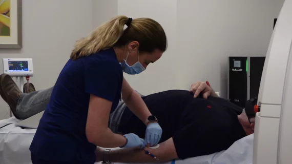
A CT coronary calcium scoring exam at Northwestern Medicine's Central DuPage Hospital in the Chicago Suburbs. Dave Fornell

The remaining gaps in evidence for cardiac CT

While the ACC/AHA 2021 Chest Pain Assessment Guidelines included cardiac CT angiography as a top level recommendation, gaps in evidence still need to be filled.

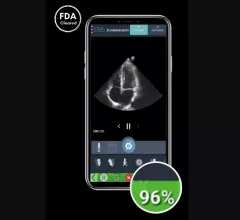
DiA Imaging Analysis, an Israel-based healthcare technology company, has gained U.S. Food and Drug Administration (FDA) clearance for LVivo IQS, a new software solution designed to help users acquire high-quality echocardiography images.

FDA clears new AI-powered cardiac imaging solution

The newly approved software uses artificial intelligence to provide users with real-time feedback related to image quality.

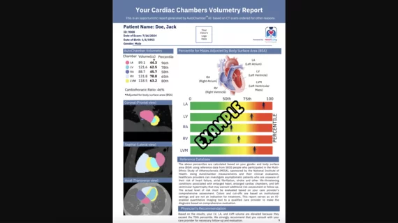
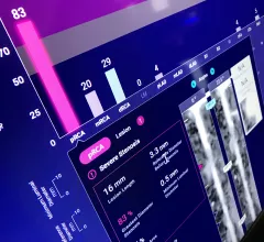
An example of an FDA cleared radiology AI algorithm to automatically take a cardiac CT scan and identify, contour and quantify soft plaque in the coronary arteries. The Cleerly software then generates an automated report with images, measurements and a risk assessment for the patient. This type of quantification is too time consuming and complex for human readers to bother with, but AI assisted reports like this may become a new normal over the next decade. Example from Cleerly Imaging at SCCT 2022.

Legal considerations for artificial intelligence in radiology and cardiology

There are now more than 520 FDA-cleared AI algorithms and the majority are for radiology and cardiology, raising the question of who is liable if the AI gets something wrong.

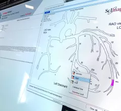
Example of a cardiovascular information system (CVIS) cath lab reporting module with a coronary tree model that will auto complete sections of the report based on how the cardiologist modifies the model. Image from the ScImage booth at ACC 2022. Photo by Dave Fornell

VIDEO: 4 key trends in cardiovascular information systems, according to Signify Reseach

Signify Research shares the latest big trends in cardiovascular IT systems, including the role of EMR cardiology modules vs. third-party CVIS, structured reporting, integration into enterprise imaging and inclusion of ambulatory surgical centers. 

#CTA #photoncountingCT #aortaCT

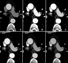
Photon counting cuts CTA contrast dosage 25%

Not only does the low-volume contrast protocol preserve supplies, it also protects patients who might be vulnerable to adverse reactions and/or side effects from contrast use. 

Thumbnail

Cardiologists use video-based AI model to ID coronary artery disease

A team of specialists out of Cedars-Sinai developed the deep learning model using TTEs from nearly 3,000 patients.

Around the web

Positron, a New York-based nuclear imaging company, will now provide Upbeat Cardiology Solutions with advanced PET/CT systems and services. 

The nuclear imaging isotope shortage of molybdenum-99 may be over now that the sidelined reactor is restarting. ASNC's president says PET and new SPECT technologies helped cardiac imaging labs better weather the storm.

CMS has more than doubled the CCTA payment rate from $175 to $357.13. The move, expected to have a significant impact on the utilization of cardiac CT, received immediate praise from imaging specialists.

Trimed Popup
Trimed Popup