Cardiac Imaging

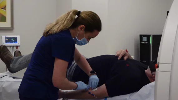
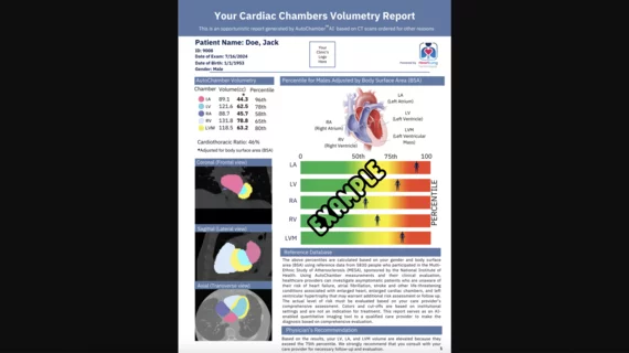
While cardiac ultrasound is the widely used imaging modality for heart assessments, computed tomography (CT), magnetic resonance imaging (MRI) and nuclear imaging are also used and are often complimentary, each offering specific details about the heart other modalities cannot. For this reason the clinical question being asked often determines the imaging test that will be used.

MITA looks to lawmakers for full repeal of medical device excise tax

The Medical Imaging & Technology Alliance (MITA) urged lawmakers in Washington, D.C., to pass a permanent, full repeal of the medical device tax.

Thumbnail

Active surveillance crucial to delaying surgery for low-risk papillary thyroid cancer

Active surveillance using ultrasonography could be a key to delaying surgical treatment for low-risk papillary thyroid cancer (PTC)—the most common type of thyroid cancer.

Left ventricular puncture proposed for whole-body PMCTA

A team of researchers has introduced a whole-body, post-mortem CT angiography (PMCTA) approach that could save time and money without requiring specialized equipment. By puncturing the left ventricle percutaneously and adding a contrast agent, researchers were able to identify threats for thromboembolism and aneurysm in a small sampling of cadavers.

PET outperforms SPECT, CCTA in diagnosing coronary artery disease

Researchers have directly compared the performance of three noninvasive imaging methods for diagnosing myocardial ischemia—and they identified positron emission tomography (PET) as the most accurate.

Trabeculation, myocardial function linked via cardiac MRI

Excess greater left ventricle (LV) trabeculation is associated with decreased average regional myocardial function, measured by myocardial strain. A team of researchers, examining MRIs of 1,123 individuals, found that average regional LV function was worse in those with greater LV trabeculation.

Thumbnail

Ground Control to Dr. Tom: Patient Exploration in an Era of Space-Age Radiology

Sponsored by Philips Healthcare

Being a radiologist today can feel a bit like being on the Starship Enterprise: you have all these Star-Trek-like tools at your disposal – devices and applications with the ability to produce incredibly sophisticated digital images and insights that we couldn’t have imagined even twenty years ago. 

Thumbnail

Prenatal 3D ultrasound shows heart defects affect brain development

European researchers have used 3D ultrasound to measure cortical folding, which increases the surface area of the brain and therefore its processing power, in fetuses with congenital heart defects. Their primary interest was in finding out whether prenatal cardiovascular anomalies affect neurodevelopment, although they also wanted to see if measuring the depths of the crevices reliably indicates the extent of folding.

Thumbnail

Cardiac MRI feasible throughout slow-to-fast ‘bike rides’

European researchers have shown that heart imagers can have their MRI and their patients on exercise bikes—pedaling harder and harder—too.

Around the web

Positron, a New York-based nuclear imaging company, will now provide Upbeat Cardiology Solutions with advanced PET/CT systems and services. 

The nuclear imaging isotope shortage of molybdenum-99 may be over now that the sidelined reactor is restarting. ASNC's president says PET and new SPECT technologies helped cardiac imaging labs better weather the storm.

CMS has more than doubled the CCTA payment rate from $175 to $357.13. The move, expected to have a significant impact on the utilization of cardiac CT, received immediate praise from imaging specialists.