Pulmonary embolism

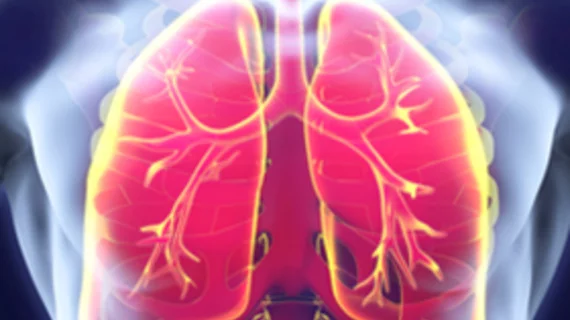
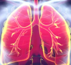
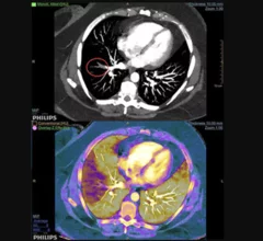
Pulmonary embolism (PE) is the third leading cardiovascular cause of death after heart attacks and stroke. PE is caused by blood clots in the pulmonary arteries. These are often caused by clots from the venous system, including thrombus from trauma, surgery or deep vein thrombosis (DVT). Treatment has traditionally been systemic use of thrombolytic drugs to dissolve the clot. But in cases there is a massive, life-threatening PE, or chronic clot burden that have remained in a vessel for an extended period of time, mechanical thrombectomy and ultrasound-assisted catheter-directed thrombolysis (USCDT) is being used as more targeted and aggressive treatments.

MRA for pulmonary embolus

MR angiography a suitable alternative to CT when ruling out pulmonary embolus

The modality switch became especially important during the iodinated contrast shortage of 2022 when clinics were forced to deploy mitigation tactics as a means of preserving their contrast supply. 

Thumbnail

VIDEO: Cardiology getting more involved in pulmonary embolism response teams

Terry Bowers, MD, director of vascular medicine at Beaumont Hospital and national co-chair of the Pulmonary Embolism Research Collaborative (PERC), explains the trend toward creating pulmonary embolism (PE) response teams that include cardiology. 

chest pain lung pulmonary embolism

Image quality is not an issue for AI model that detects pulmonary embolisms on CT

CTPA is the standard of care for diagnosing PE, but suboptimal scans make it difficult to reach a diagnosis. A new Clinical Imaging study tests the effectiveness of AI when image quality is lacking.

Thumbnail

Negative CT-PA scans could increase time-to-disposition in patients seen for pulmonary embolism

Patient and environmental factors proved not to be major culprits but scan results were key, experts reported in BMC Emergency Medicine.

Thumbnail

Chest CT, perfusion imaging spots pulmonary embolism in 25% of hospitalized COVID patients

Men and smokers were particularly prone to this condition, which, if left untreated, carries an up to 30% mortality rate.

chest pain lung pulmonary embolism

New pulmonary embolism approach could substantially reduce imaging overuse

The pretest probability score produced false-negative rates below 1% and dropped imaging use by about 20%, according to a new JAMA Cardiology study. 

chest pain lung pulmonary embolism

AI spots dozens of missed incidental pulmonary embolism diagnoses at one hospital

The investigation was retrospective, but Duke scientists believe their algorithm could potentially aid radiologists in spotting near-misses in their work.

Example of spectral, or dual-energy CT, confirming a pulmonary embolism (PE). Image courtesy of Philips Healthcare

Dual-energy CTA iodine maps offer small benefit in diagnosing pulmonary embolism

Dual-energy (DE) CT iodine maps offer a slight benefit when paired with traditional CT angiography images in diagnosing pulmonary embolism (PE), reported researchers in a Sept. 11 Radiology study.

Around the web

In the post-COVID era, wages for permanent RNs are rising, and wages for travelers are decreasing. A new report tracked these trends and more. 

Debra L. Monticciolo, MD, past president of both the Society of Breast Imaging and the American College of Radiology, explains the advantages and disadvantages of current breast screening technology.

The new guideline details the best imaging strategies for a variety of clinical scenarios. 

Trimed Popup
Trimed Popup