Chest x-ray may overlook coronavirus cases apparent on CT
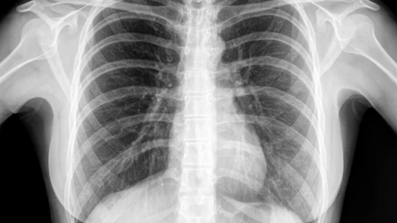
A “considerable” number of patients with COVID-19 did not have abnormalities on their chest x-rays suggestive of the virus, according to a multi-center study published in the Korean Journal of Radiology. The findings indicate the modality may miss critical cases of this growing epidemic.
Researchers retrospectively looked at the x-ray and CT findings from a handful of patients with coronavirus pneumonia to draw their conclusions. Most of the abnormalities reviewed on chest CT scans were congruent with prior academic findings, but only 33% of patients had abnormal radiographic findings, the authors wrote Feb. 26. Although preliminary, radiologists should turn to computed tomography when evaluating suspected coronavirus cases.
“Most of the pulmonary lesions were ambiguous on chest radiographs,” Soon Ho Yoon, MD, PhD, with Seoul National University Hospital in South Korea, and colleagues wrote. “Clinicians and radiologists should become familiar with the CT findings of COVID-19 and the limitations of chest radiographs in evaluating pneumonia to manage the COVID-19 outbreak.”
For the study, Yoon and colleagues from four other Korean healthcare institutions analyzed the imaging results of nine patients. They found chest radiography fell woefully short of CT in picking out infected individuals.
In total, three patients had parenchymal abnormalities detected by chest radiography, and a majority of these were peripheral consolidations. Chest CT discovered “bilateral involvement” in all but one patient. These lesions were often ill-defined and made up of mixed ground-glass opacities and consolidations or pure ground-glass opacities.
Yoon and colleagues also pointed out that COVID-19 yields CT abnormalities that are similar to those produced by severe acute respiratory syndrome (SARS) and Middle East respiratory syndrome (MERS)—both of which are part of the coronaviridae family. However, coronavirus appears to be “radiologically milder” than both viruses, they added.
The researchers acknowledged that their sample size was less than ideal and represented only one-third of the 29 identified patients with coronavirus in Korea during the time of their study.
The study is the latest of several to confirm CT’s superiority in detecting individuals with the virus that has claimed 3,254 lives and sickened more than 95,000. Its spread into Europe has also forced major healthcare societies to delay their conferences. Just yesterday, March 3, the European Society of Radiology announced it was postponing its annual meeting by nearly four months because of the outbreak.

