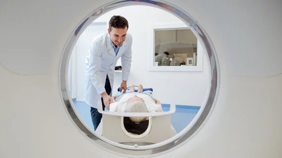
MRI helps diagnose central nervous system tumors
MRI can predict the severity of one of the most frequent tumors of the central nervous system, according to a recent study published in Clinical Radiology. Researchers believe it may help tailor a patient’s management plan.
“Although mostly benign, meningiomas still represent a major challenge to neurosurgeons and other medical disciplines involved in their diagnostic and therapeutic management,” wrote F. Salah, American University of Beirut Medical Center in Lebanon, and colleagues. “As modern techniques have greatly improved the prognosis of meningioma patients, the value of clear grading criteria for meningiomas via brain imaging is important to determine the best management protocol and reduce the risk of complications during and after surgery.”
The researchers sought to compare features on both quantitative and qualitative MRI and CT for differentiating benign from malignant meningiomas. To do so, they evaluated scan features of 71 patients who underwent surgical resection of intracranial meningiomas at American University between 2008 and 2017.
Overall, MRI showed great promise for detecting meningioma brain invasion. It scored a specificity of 79%, negative predictive value of 92%, positive predictive value of 7% and sensitivity of 20%.
Upon univariate analysis, multiple MRI features correlated to high-grade meningiomas, including tumor size and volume, heterogenous enhancement, presence of intra-tumoral necrosis and brain invasion. On CT, hyperostosis of the adjacent skull was the only predictor of low-grade meningioma.
“The results of the present study showed that the most significant findings to differentiate between the two groups included a heterogeneous pattern of enhancement, as well as the presence of intra-tumoral cystic change or necrosis; both more described in GP2 meningiomas,” the authors wrote. “This can further explain that both imaging features are correlated.”
Salah and colleagues noted their study was strengthened by the variety of CT and MRI features included, as well as their choice to quantify the volume of oedema instead of grading it subjectively.
They also suggested a radiomics feature-based machine learning classifier would be “interesting” to test, but is not widely available for all radiologists.
“MRI has a promising role in predicting meningioma grade prior to resection, which can directly impact future management protocols regarding surgical planning and complications,” the researchers concluded.

