Imaging Informatics

Imaging informatics (also known as radiology informatics, a component of wider medical or healthcare informatics) includes systems to transfer images and radiology data between radiologists, referring physicians, patients and the entire enterprise. This includes picture archiving and communication systems (PACS), wider enterprise image systems, radiology information. systems (RIS), connections to share data with the electronic medical record (EMR), and software to enable advanced visualization, reporting, artificial intelligence (AI) applications, analytics, exam ordering, clinical decision support, dictation, and remote image sharing and viewing systems.

Example of natural language processing converting the radiologist's dictation into text. This system from M-Model highlighted key words the artificial intelligence will use text in the report and for labeling the report file for later key word searches or data mining. 

How NLP can 'revolutionize' structured reporting

The continued emergence of natural language processing has caught the eye of experts in the field, with some suggesting its use could streamline the process of integrating structured reporting across the specialty. 

Traditional methods continue to outperform AI in some orthopedic scenarios

A new meta-analysis suggests that when it comes to hip fractures, AI algorithms do not always live up to their hype. 

Russian-speaking ransomware group leaks breast cancer patients' sensitive data

"We have been in your network a long time and have had time to study your business. We have stolen your confidential information and are ready to publish it," the ransomware group threatened.

Rankings of radiology IT solutions by end-users in the 2023 Best in KLAS program

End-users of various radiology IT systems offer their assessment of the software in the annual KLAS Research 2023 Best in KLAS report.

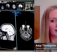
The integration of artificial intelligence (AI) into radiology PACS and enterprise imaging systems has become a big topic of discussion with IT vendors over the past couple years. This has become a bigger question from hospitals and radiology groups as there are now about 400 radiology related AI algorithms that have U.S. Food and Drug Administration (FDA) clearance. Amy Thompson, a senior analyst at Signify Research, is monitoring AI trends in radiology and discusses trends.

Trends in the adoption and integration of AI into radiology workflows

Amy Thompson, a senior analyst at Signify Research, explains why AI adoption has been slow in radiology, common barriers and trends in the market.

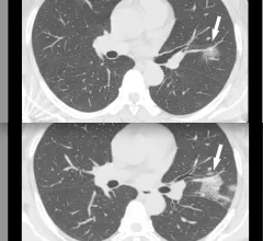
CT of coronavirus pneumonia, a solitary rounded ground-glass opacity (GGO) pattern. A 51-year-old woman in China presented in January 2020 without fever, but had close contact with positive patients. Top, baseline axial unenhanced chest CT obtained 6 days before the first positive PCR test. Bottom, chest CT scan 4 days later shows the size increase of the lesion (arrow). Image courtesy of RSNA. #COVID #SARSCoV2

How effective are chest CT severity scores in managing COVID?

While the studies on these systems have proven them to be effective in diagnosing and treating COVID in specific cohorts, the varying settings in which they were used can make it difficult to derive definitive conclusions on their efficacy.

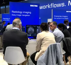
Amy Thompson, a senior analyst at Signify Research, explains what she is seeing in the market for radiology PACS. She said the biggest overall, strategic technology trends are wider adoption of enterprise imaging systems expanding beyond radiology to include other departments, migration to cloud data storage, and adoption of artificial intelligence. Components of these integrate into the 5 trends in radiology IT systems outlined below.

5 key trends in PACS and enterprise imaging from Signify Research

Signify Research explains several key trends in the evolution of radiology PACS and enterprise imaging systems, including adoption of artificial intelligence, streamlining workflow, implementing structured reporting and more.

long covid lung CT

Some long COVID patients continue to display multi-organ damage one year after recovery

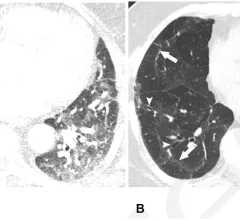
A new study utilizing multi-organ MRI scans recently identified organ impairment in 62% of COVID long haulers six months after their initial diagnosis; 29% of these individuals continued to display damage in at least one organ at the 12-month mark.

Around the web

The nuclear imaging isotope shortage of molybdenum-99 may be over now that the sidelined reactor is restarting. ASNC's president says PET and new SPECT technologies helped cardiac imaging labs better weather the storm.

CMS has more than doubled the CCTA payment rate from $175 to $357.13. The move, expected to have a significant impact on the utilization of cardiac CT, received immediate praise from imaging specialists.

The newly cleared offering, AutoChamber, was designed with opportunistic screening in mind. It can evaluate many different kinds of CT images, including those originally gathered to screen patients for lung cancer. 

Trimed Popup
Trimed Popup