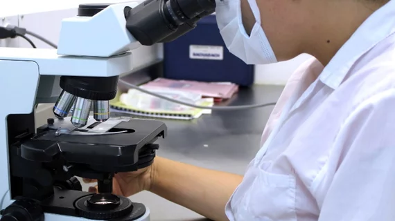
Open-source tool ensures quality-control for digital pathology slides
Case Western Reserve University researchers have released a new tool to ensure the quality of digital pathology images being used for diagnostic and research purposes.
HistoQC, created by bioengineering researchers in Case Western’s Center for Computational Imaging and Personal Diagnostics, is an open-source quality-control tool that helps users flag low-quality images while preserving those that can help clinicians make accurate diagnoses.
"The idea is simple: assess digital images and determine which slides are worthy for analysis by a computer and which are not," said Anant Madabhushi, professor of biomedical engineering at the Case School of Engineering, in a news release. "This is important right now as digital pathology is taking off worldwide and laying the groundwork for more use of AI (artificial intelligence) for interrogating tissue images."
Currently, there are no go-to standards for preparing and digitizing pathology slides, low-quality images often are mixed in with higher-quality slides.
This was evident when Andrew Janowczyk, co-creator of HistoQC, analyzed slides housed in the Cancer Genome Atlas, a collection of more than 30,000 cancer tissue samples. Nearly 800 slides, or 10% had problems, consisting of a crack or air bubbles between the slide’s layers.
The new tool could be a step in ensuring the highest quality slides for digital pathology workflows.
“… HistoQC could provide an automated, quantifiable, quality control process for identifying artefacts and measuring slide quality, in turn helping to improve both the repeatability and robustness of DP workflows,” the researchers wrote.
Research for the HistoQC tool is supported by a three-year, $1.2 million grant from the National Cancer Institute. The open-source tool was published in the Journal of Clinical Oncology Clinical Informatics.

