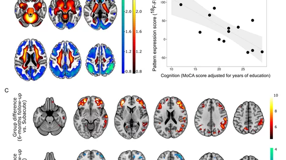
PET scan depicting neurological fallout of COVID-19 wins image of the year
For patients suffering from neurological side effects of COVID-19, positron emission tomography can play a crucial role in measuring neural impairment.
The findings come from a study of individuals recently diagnosed with the novel virus who underwent inpatient treatment and brain imaging. PET scans depicted areas of cognitive impairment, neurological symptoms and disease progression over a six-month time period.
The Society of Nuclear Medicine and Molecular Imaging selected this brain scan (pictured above) as the Henry N. Wagner Jr. Image of the Year late Tuesday during the organization’s annual meeting. The image beat out more than 1,280 abstracts to take home the prize.
“18F-FDG PET is an established biomarker of neuronal function and neuronal injury,” SNMMI’s Scientific Program Committee Chair, Umar Mahmood, MD, PhD, said in a statement. “As shown [in] the Image of the Year, it can be applied to unravel neuronal correlates of the cognitive decline in patients after COVID-19.”
To achieve their award-winning results, the authors prospectively studied COVID patients admitted to the hospital for non-neurological complaints. Participants underwent a cognitive assessment and, if at least two neurological symptoms were apparent, also received a PET scan.
Compared to healthy patients, individuals with COVID-19 showed distinct brain metabolism patterns. Those included marked metabolic decreases in cortical regions and patterns consistent with patients’ cognitive functioning.
There was also good news, the team reported. Upon six-month follow-up, PET imaging showed neurological improvement across most patients, including a near full recovery of brain metabolism.
Ganna Blazhenets, PhD, a post-doctoral researcher in Medical Imaging at the University Medical Center Freiburg in Germany, noted their research will be important as post-COVID problems continue to emerge.
“We can clearly state that a significant recovery of regional neuronal function and cognition occurs for most COVID-19 patients based on the results of this study. However, it is important to recognize the evidence of longer-lasting deficits … in some patients six months after manifestation of disease,” Blazhenets added. “As a result, post-COVID-19 patients with persistent cognitive complaints should be presented to a neurologist and possibly allocated to cognitive rehabilitation programs.”
You can read the abstract for this study here.

