Radiologists use diagnostic imaging to non-invasively look inside the body to help determine the causes of an injury or an illness, and confirm a diagnosis. Providers use many imaging modalities to do so, including CT, MRI, X-ray, Ultrasound, PET and more.
Following a recent surge of mycoplasma pneumoniae pneumonia cases, experts have issued new guidance to help providers quickly identify and treat the condition, with imaging playing a prominent role.
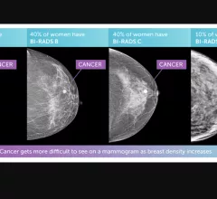
An artificial intelligence system that is currently commercially available for use in adults could also have applications in a pediatric population, according to a new study in Pediatric Radiology.
Munir Ghesani, MD, President of the Society of Nuclear Medicine and Molecular Imaging (SNMMI), system chief of nuclear medicine at Mount Sinai Health, explains recent advances in nuclear imaging technology.
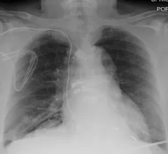
After experts from one institution evaluated 500 portable chest x-rays completed during the summer of 2021, it was revealed that 46.2% of the images obtained were problematic, requiring the imaging to be repeated.
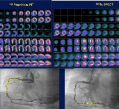
The biggest news from the American Society of Nuclear Cardiology (ASNC) 2022 meeting was positive late-breaking data on the phase 3 Aurora trial for the flurpiridaz (F-18) PET radiotracer agent.
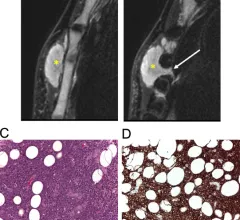
A new analysis offers a detailed comparison of soft-tissue lymphomas and soft-tissue tumors based on imaging characteristics from MRI scans—an area of study that has not yet been rigorously explored, the authors of the paper indicated.
When combined with artificial intelligence-based noise reduction techniques, new photon-counting CT technology can increase the detection of bone disease while also decreasing radiation exposure.
Clinicians have been using HeartSee to diagnose and treat coronary artery disease since the technology first debuted back in 2018. These latest updates, set to roll out to existing users, are designed to improve diagnostic performance and user access.
The cardiac technologies clinicians use for CVD evaluations have changed significantly in recent years, according to a new analysis of CMS data. While some modalities are on the rise, others are being utilized much less than ever before.