
Some imaging features of missed lung cancer indicate worse outcomes
Certain radiographic features of missed lung cancer (MLC) could be indicative of worse outcomes for patients, according to new research.
Features pertaining to location, density and superimposed structures were all found to be associated with poorer outcomes for patients who initially had their lung cancer overlooked on radiographs, a new paper in Clinical Imaging recently revealed.
“Because MLC tends to grow over time and progress from early stage, potentially curable LC to advanced-stage LC, failure to identify or describe an incidentally encountered abnormality subsequently proven to be LC on a chest radiograph can lead to poor patient outcomes, medicolegal issues, or malpractice litigation,” corresponding author of the paper Thitiporn Suwatanapongched, with the Division of Diagnostic Radiology at Faculty of Medicine Ramathibodi Hospital, and colleagues explained.
After retrospectively reviewing the cases of 95 patients who received chest X-rays at least six months prior to their lung cancer diagnosis, the team identified several radiographic features associated with advanced stage cancer and poor outcomes.
The following cancer imaging features were found to have a significant association with stage 3 or higher initial lung cancer diagnosis:
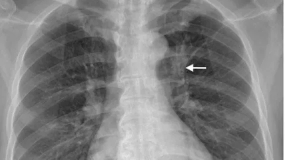
Being located in the inner one-third of the lung.
Having a density greater than or equal to the aortic knob.
Being superimposed by midline structures (hilum, heart, thoracic aorta).
Specifically, three-year all-cause mortality significantly increased if the missed cancer was located in the upper zone, superimposed by pulmonary vessels, superimposed by pulmonary vessels plus ribs, or was superimposed by pulmonary vessels while also being located in the inner one-third of the lung.
The team suggested that their findings pertaining to density could reflect the solid, more invasive behavior of lung cancer, while the anatomy in the inner one-third of the lung that includes superimposing anatomical structures could make it difficult to detect tumors in their earlier stages.
“These factors may contribute to a larger tumor size or mediastinal structure involvement at diagnosis, which can preclude curative surgical treatment,” the group noted.
When radiologists come across these findings incidentally, the authors suggested that “special attention and prompt reporting to clinicians for follow-up or referral for additional imaging” is warranted.
The study abstract is available here.

In addition to her background in journalism, Hannah also has patient-facing experience in clinical settings, having spent more than 12 years working as a registered rad tech. She began covering the medical imaging industry for Innovate Healthcare in 2021.