NASA astronauts use ultrasound in space to evaluate spinal health
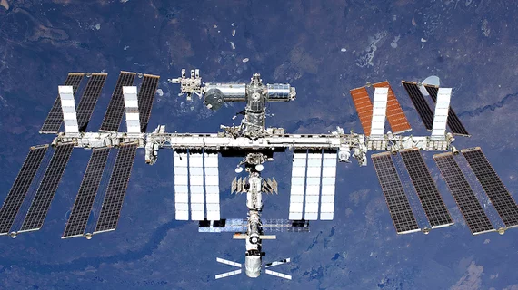
While at the International Space Station (ISS), astronauts used portable ultrasound technology to scan each for spinal cord changes that may occur during long-term space missions and cause prolonged back pain, according to a study published in the April issue of the Journal of Ultrasound in Medicine.
The astronauts were able to obtain diagnostic quality spinal images with a 92.5 percent success rate, according to study researchers led by Kathleen Garcia, BSc from Henry Ford Hospital in Detroit.
"Focused ultrasound monitoring of the spine for longitudinal changes during long-duration spaceflight may influence additional strategies or nutrition/drug therapies to reduce disk degeneration," Garcia et al. Wrote. "Given the previous void of in-flight spinal imaging capabilities in space, to our knowledge, this study represents the first attempt to monitor microgravity-associated acute changes to the spine while they are occurring."
The researchers worked with NASA to train nine astronauts (eight men with a mean age of 46 years old) scheduled for a long duration flight on the ISS to perform spinal ultrasound examinations on each other. The astronauts were trained six months before their mission.
A total of seven astronauts were trained for ultrasound operator and research patient roles, with the other two trained as backup operators. The seven astronauts who provided consent to be research patients underwent lumbar and cervical spinal ultrasound exams before flight, in flight and after flight.
This data were then compared to pre- and post-flight MRI.
Image acquisition success rates were 95 percent in the lumbar spine and 90 percent in the cervical spine, along with "no appreciable difference in success rates for either image acquisition or image quality between expert operators and astronaut crew members in the lumbar and cervical regions," the researchers wrote.
Preflight examinations revealed 33 spinal findings, with an additional 14 spinal findings identified from post-flight scans. Disk desiccation, osteophytes and qualitative changes in the intervertebral disk height and angle were found.
Additionally, more than half of the astronauts reported having back and neck pain during early spaceflight and after returning.
Overall, although the researchers acknowledged the limitations of a small sample size, they hope their research may provide further insight into the pathophysiologic mechanisms of space-related back pain with ultrasound being a viable tool for astronauts to collect accurate and quality imaging data during long-term space flights.
"As the duration of space missions continues to increase, this new utility will only gain importance in monitoring crew health and diagnosing disorders," the researchers wrote. "Further investigations should be performed to corroborate this imaging technique and to create a larger database related to in-flight spinal disorders during long-duration spaceflights, and efforts should be made to incorporate these findings into the development and refinement of microgravity countermeasures."

