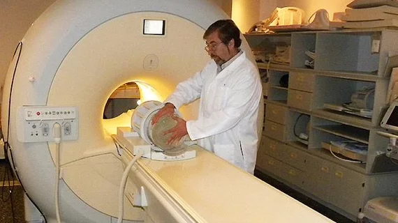
Novel MRI method measures myelin in MS, stroke, TBI patients to evaluate therapy, recovery
A novel brain MRI based method can track myelin— responsible for insulating nerve fibers — and may enable clinicians to identify myelin content changes in MS patients and patients whose myelin has been damaged by stroke or traumatic brain injury.
The method is currently being developed by Vasily Yarnykh, PhD, a radiology researcher and associate professor at the University of Washington School of Medicine, who has developed myelin imaging protocols to work with MRI scanners from numerous vendors in U.S. hospitals, as reported by the University of Washington School of Medicine.
“For instance, with a stroke there is a chemical death of brain tissue–neurons, glial cells–and disruption of myelin,” Yarnykh said in a prepared statement. “As neurons recover, axons start to grow and establish new neural connections. There’s the question of how to measure these competing processes of demyelination and remyelination as the brain tries to heal.”
In MS patients, Yarnykh explained, myelin levels could indicate the effectiveness of therapies or general neural tissue recovery in stroke or brain injury patients. There’s also the potential to measure brain development in infants.
Two weeks ago, Yarnykh won a National Institutes of Health High-Impact Neuroscience Research Resource award of $700,000 to make the myelin-measurement technology available to the scientific and medical community over the next four years.
“In addition to the imaging protocols themselves, we need to put this method in the hands of end-user MRI technicians, so the software needs to be user-friendly and downloadable,” Yarnkyh said. “We’ll have to run quality-assurance procedures and provide some test objects to show that the method works correctly on each MRI machine.”

