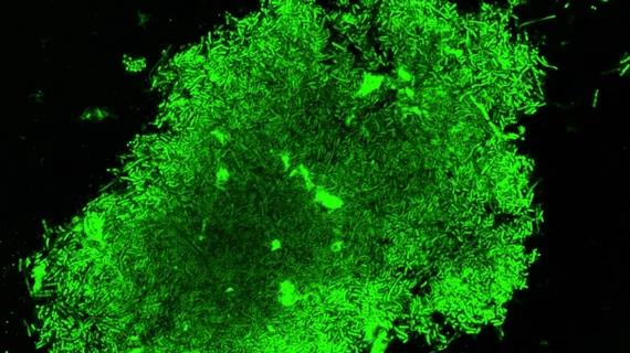
Stanford’s 'glowing' imaging technique could diagnose TB in an hour
Tuberculosis (TB) could now be diagnosed in as little as an hour with a new imaging technique guided by glowing bacteria and developed by researchers at Stanford University. Their research was published online on Aug. 15 in Science Translational Medicine.
As many TB strains have become defiant against or immune to standard treatments, there is a growing need for quicker, more cost-effective TB diagnostic methods to decrease infection rates. Because the new imaging method could be cheaper and quicker, hospitals in developing countries where TB is most prominent could greatly benefit from it, according to the researchers, led by Jianghong Rao, PhD, a professor of radiology at Stanford.
The imaging technique requires a standard fluorescence microscope and a spit sample from the patient.
When the two-piece fluorescent probe encounters live TB bacteria, one part of the probe activates, or glows green, and the other localizes the glow of the concentrated fluorescence to the bacterium, allowing the researchers to see and track the infection.
"For cases of drug-susceptible TB, the treatment success rates are at least 85 percent, but the rate of success is only 54 percent for multidrug-resistant TB, which requires longer treatments and more expensive, more toxic drugs," Rao said in a prepared statement.
The researchers explained the fluorescent probe can help determine the appropriate drug by showing which bacteria are still alive in the patient sample. In the future, they hope their technique could help develop new TB drugs and determine which drugs work best to combat individual strains of TB.
"The hope is to make this an adaptable technology," Rao said. "It's something that could be really widespread, and you wouldn't necessarily need to use it in a hospital setting."

