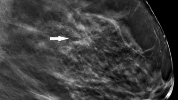
Tomosynthesis trumps mammo alone for spotting architectural distortion
Digital breast tomosynthesis (DBT), also called 3D mammography, has been shown to provide better visualization of architectural distortion, a subtle mammographic finding. It also identifies some distortions that would not be found at all if screening was performed using digital mammography alone, according to a study published in the July issue of American Journal of Roentgenology (AJR).
Architectural distortion is a distortion of the breast tissue with no definite visible mass, including distortion at the edge of the parenchyma. It’s the third most common mammographic manifestation of nonpalpable breast cancer, but is often missed at screening mammography, explained Luke Partyka, MD, of Alpert Medical School of Brown University in Providence, R.I., and colleagues.
“It has been hypothesized that [digital breast tomosynthesis] is likely to have its greatest potential benefit in cases of particularly subtle mammographic findings such as architectural distortion,” wrote the authors.
To put this hypothesis to the test, Partyka and colleagues retrospectively reviewed BI-RADS category 0 reports from nearly 10,000 screening digital mammography exams with adjunct tomosynthesis, searching for the term “architectural distortion.” These were reviewed in consensus by three radiologists, who noted whether architectural distortions were seen more easily on digital mammography, tomosynthesis or equally as well on both.
Twenty-six cases of architectural distortion were identified in the review, 19 (73 percent) of which were seen only on digital breast tomosynthesis images, reported Partyka and colleagues. “In these cases, although visualization on [digital mammography] was confirmed retrospectively in our consensus review, it is uncertain that the finding of [architectural distortion] would have been made prospectively without the added benefit of the tomosynthesis images calling attention to the areas in question.”
Six of the remaining seven architectural distortions were seen more clearly on tomosynthesis than digital mammography. Nine lesions were assigned to BI-RADS category 4 or 5 upon diagnostic workup. The cancer detection rate of digital breast tomosynthesis in mammographically occult architectural distortion was 21 percent.
Partyka and colleagues suggested the need to establish a guideline for the workup of architectural distortion detected on screening tomosynthesis, and that careful correlation with surgical history is needed in cases of post-surgical architectural distortion.
“Based on our initial experience, architectural distortion that is not post-surgical and is seen only on [digital breast tomosynthesis] should not be easily dismissed, and further evaluation with ultrasound should be performed.”
More Coverage of Architectural Distortion:
DBT detects more architectural distortion lesions than 2D mammography alone
When does worrisome architectural distortion signal malignancy on mammography?
New research can help radiologists manage architectural distortion identified via DBT exams
3D mammography detects more architectural distortions than 2D

