
Mammograms should not be delayed after COVID vaccine, research shows

Women who experience swollen lymph nodes after COVID vaccination should still complete their screening mammograms without delay, according to new research published Tuesday in Radiology.[1] This is a change in direction since early 2021 for breast screening, when it was discovered some women develop axillary adenopathy (swollen lymph nodes) after receiving the COVID-19 vaccine, which can be interpreted on mammograms as a sign of cancer.
Many women who had mammograms shortly after receiving a COVID vaccine were surprised to learn of their lymphadenopathy findings. As these instances became increasingly common, research suggested the abnormalities were likely benign in nature. Despite this, experts still recommended that women temporarily delay their mammograms if they had recently received the vaccine, as the swelling was expected to subside shortly thereafter.
However, continued research efforts have contradicted that notion and have shown many cases in which the lymphadenopathy lasts for weeks or months. That is what led experts to study the long-term outcomes of those who had abnormal breast imaging after vaccination.
“This is the largest study to evaluate axillary lymphadenopathy after COVID-19 vaccination, with long term follow-up data,” Stacey Wolfson, MD, with the Department of Radiology at New York University Grossman School of Medicine and NYU Langone Health in New York City, said in a statement. “This is important because it gives us further insight as to the natural behavior of this reactive lymphadenopathy and allows us to make broader guidelines regarding follow-up.”
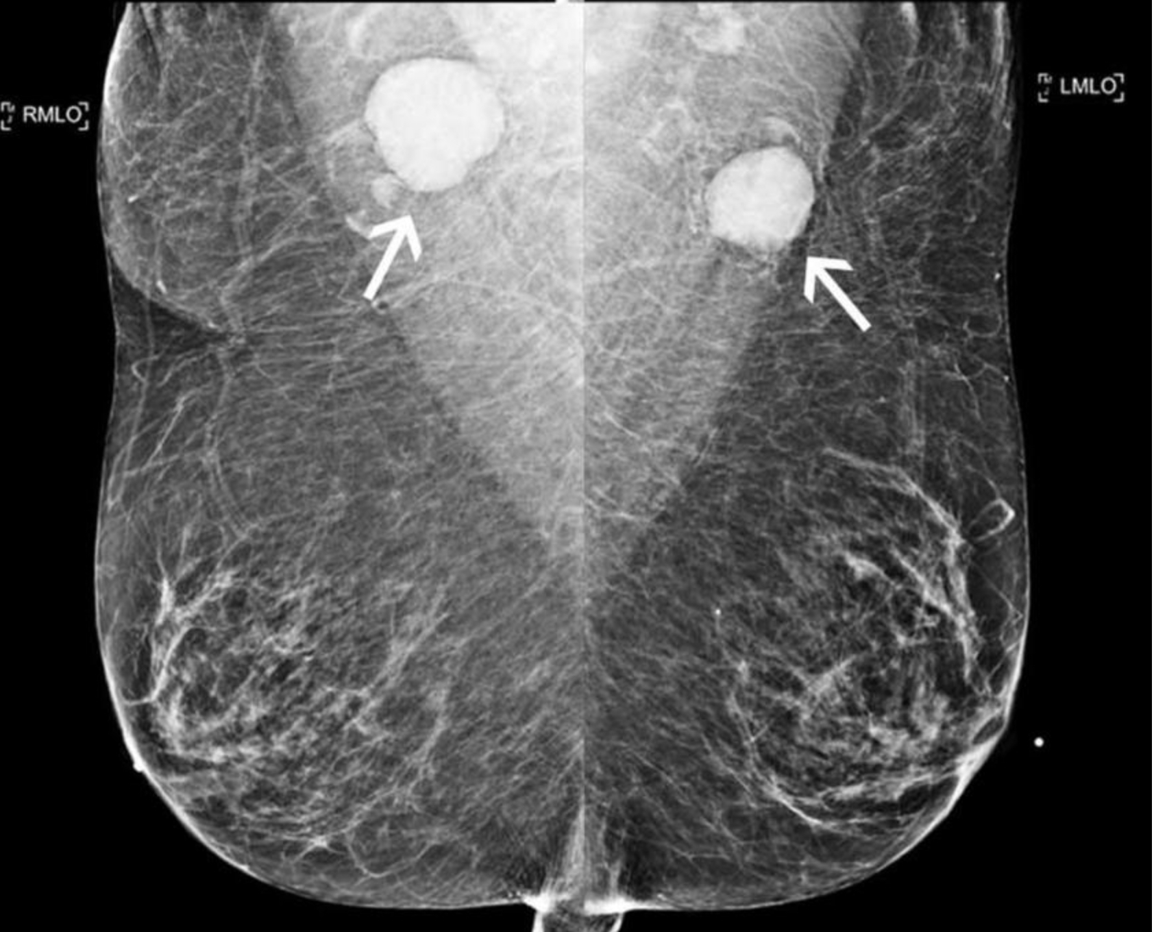
Mammography imaging showing axillary adenopathy caused by COVID-19 vaccination. The swollen lymph nodes can mimic cancer. A new study suggests women should not delay screening because of the issue. One example from the study of a 78-year-old woman following COVID-19 vaccination. Top row: Bilateral MLO views from comparison mammogram performed one year prior demonstrated morphologically normal appearing lymph nodes. Bottom row: Bilateral mediolateral oblique views following COVID-19 vaccination demonstrated new bilateral axillary lymphadenopathy with enlarged and dense lymph nodes (arrows). Axillary lymph node biopsy yielded follicular lymphoma. Image courtesy of RSNA
From December 30, 2020, to April 12, 2021, researchers pulled exam information from 17 sites within one institution. They were able to identify 1,217 women who had received a COVID vaccine, breast imaging and follow-up exams during that time.
Of those women, 44% (537) showed lymphadenopathy on at least one imaging exam (mammogram, ultrasound or both together). The swollen axillary lymph nodes developed anywhere from 1-71 days after vaccination, and in some patients the swelling persisted for up to 43 weeks after receiving their second dose.
“We found benign reactive lymph nodes were still present despite delaying the screening exams for 4 to 6 weeks based on various guidelines,” Wolfson noted. “These lymph nodes were unchanged with follow up exams at three months, and some enlarged lymph nodes persisted for over 10 months.”
The findings prompted experts to recommend that there be no delay between vaccination and screening mammograms, as reactive lymphadenopathy seen on imaging can last for several months in some cases and should be considered in conjunction with a patient’s risk factors before considering additional imaging or invasive biopsy.
Read the full article here.
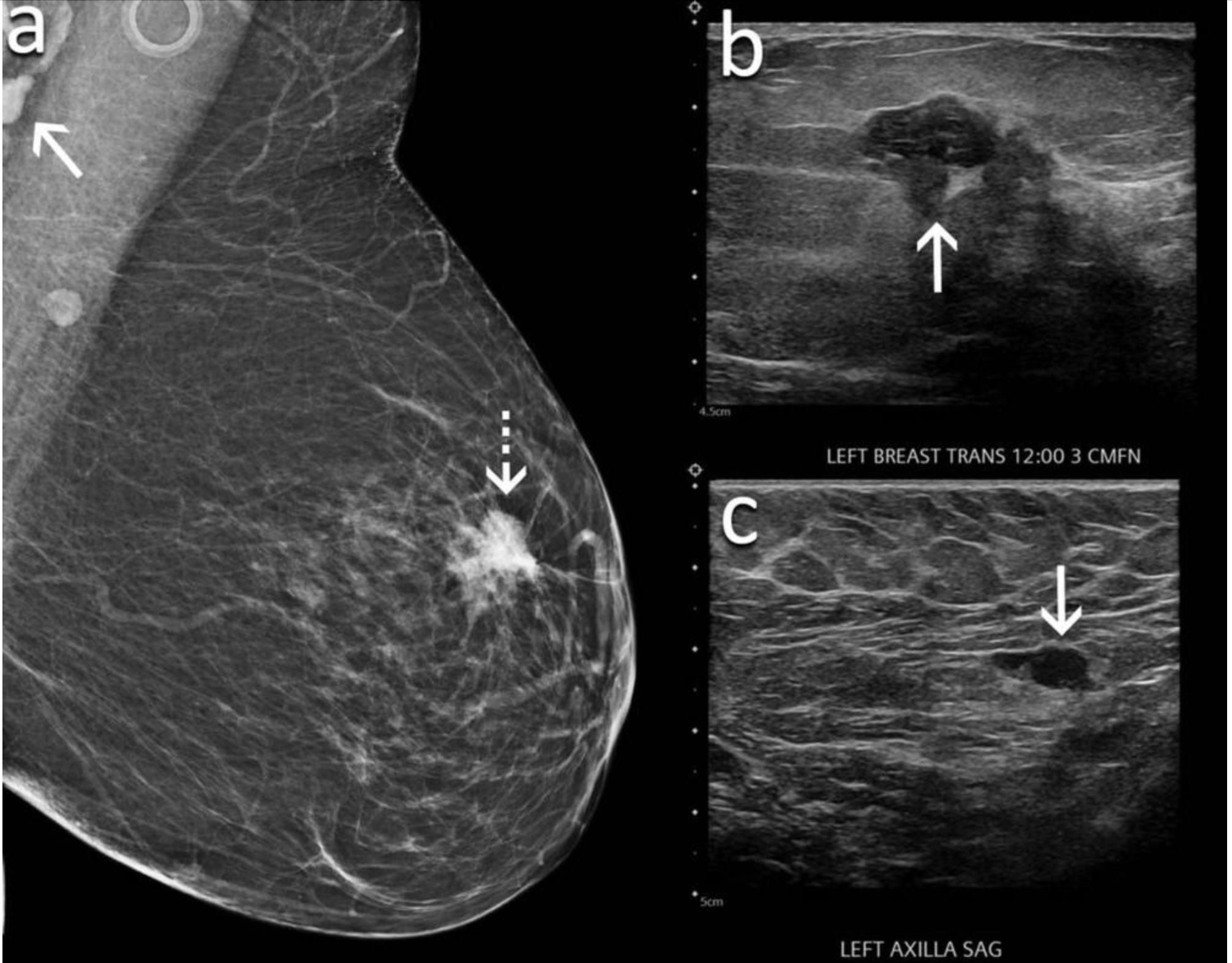
An example from the study of a 60-year-old woman presented for screening mammography nine weeks after receiving her second dose of the Moderna COVID-19 vaccine. (A) Mediolateral oblique view of the left breast demonstrates an irregular mass in the superior left breast (dash arrow) and a dense lymph node in the left axilla that is incompletely visualized (arrow). Sonography demonstrates (B) an irregular hypoechoic mass in the breast (arrow) that corresponded to the mammographic finding and (C) a lymph node with eccentric cortical thickening measuring 6 mm (arrow) in the axilla. Biopsy of the mass yielded invasive ductal carcinoma and biopsy of the lymph node yielded metastatic carcinoma. Image courtesy of RSNA
Related COVID vaccine-induced adenopathy in breast imaging:
Q&A: Should COVID vaccinated patients delay getting breast imaging — new study says no
How radiology providers can minimize mammography recalls among COVID-vaccinated patients
Most routine imaging should be scheduled 6 weeks after 2nd vaccine dose, major cancer centers say
Managing COVID-19 vaccine side effects: Harvard radiologists share their ‘pragmatic’ approach
COVID-19 vaccine update: Radiologists report side effects mimicking breast cancer on mammograms
Reference:

In addition to her background in journalism, Hannah also has patient-facing experience in clinical settings, having spent more than 12 years working as a registered rad tech. She began covering the medical imaging industry for Innovate Healthcare in 2021.