CT scans reveal 9/11 responders face increased risk of liver disease
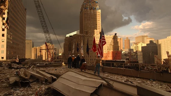
Responders who arrived at the World Trade Center complex immediately after the 9/11 attacks face a higher likelihood of developing liver disease compared to those who worked later in the rescue and during recovery efforts, according to new imaging data.
Mount Sinai experts based their conclusions on more than 1,700 lung CT scans included in the New York health system’s World Trade Center Health Program Clinical Center of Excellence. Those who arrived at ground zero within about two weeks of the attack had more exposure to toxic dust and more evidence of liver disease on their exams.
The health program monitors more than 20,000 responders who were unprotected from toxicities during the aftermath of September 11, 2001. And this research suggests clinicians should maintain a watchful eye for signs of hepatic steatosis in these individuals.
“Our study showed that continued monitoring for liver disease is warranted in World Trade Center responders…particularly those who arrived at or shortly after the attacks and had a higher exposure to the toxic dust,” senior author Claudia Henschke, MD, PhD, professor of Diagnostic, Molecular and Interventional Radiology at Mount Sinai’s Icahn School of Medicine, said in a statement. “At the moment, there are no protocols to monitor responders for liver disease, so this study points to the need to further study this issue in this at-risk population.”
Henschke et al. utilized an automated algorithm to estimate CT liver density in first responders. Lower density measures—evidence of fatty liver disease—were detected in nearly 14% of participants. Those who arrived on the scene earlier showed more evidence of disease on their scans.
“Our previous work found evidence of liver disease was three times higher in the lung scans of World Trade Center responders compared to other patients’ lung scans, so this new study suggests that responders who arrived at Ground Zero earlier should receive enhanced monitoring for liver disease,” Artit Jirapatnakul, PhD, assistant professor of Diagnostic, Molecular and Interventional Radiology explained. “Now that we have this link, the next step is to understand why or how the toxic dust actually causes liver damage.”
Read the full study published in the American Journal of Industrial Medicine here.

