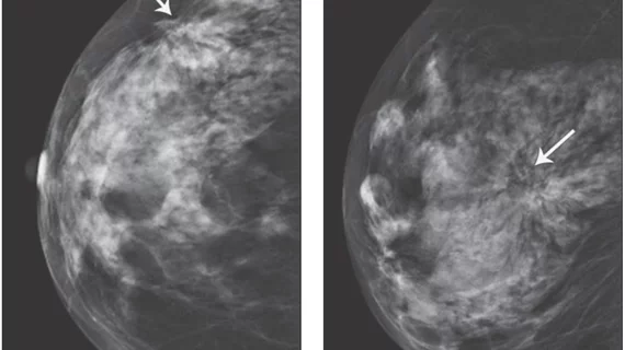
When does worrisome architectural distortion signal malignancy on mammography?
Architectural distortion, the non-mass but potentially ominous clinical feature observed in many breast imaging procedures, is less likely to signal malignancy when it is detected on screening mammography rather than diagnostic mammography or when it does not correlate with a subsequent targeted breast ultrasound exam.
In the 10-year study behind this conclusion, malignancy-suggestive architectural distortion was observed in 435 of 231,051 (0.2 percent) mammographic exams performed on women between 27 and 92 years old (mean age, 58 years).
The study, led by Manisha Bahl, MD, MPH, of Duke University, and published in the December edition of the American Journal of Roentgenology (AJR), placed the positive predictive value of architectural distortion for malignancy at 74.5 percent.
Bahl and colleagues excluded 66 cases from the 435 because these were either associated with a mass (62) or had no available pathology results (4).
Of the 369 worrisome architectural-distortion cases they retrospectively reviewed, 275 had invasive adenocarcinoma or ductal carcinoma in situ (DCIS) — hence the PPV of 74.5 percent.
Specifically, the most common pathologic finding was invasive ductal adenocarcinoma with or without lobular features (195 of 369, 52.8 percent), followed by invasive lobular adenocarcinoma with or without ductal features (64 of 369, 17.3 percent), invasive adenocarcinoma with mixed ductal and lobular features (1 of 369, 0.3 percent) and DCIS (15 of 369, 4.1 percent).
DCIS alone was identified in only 4.1 percent (15 of 369).
Watch a VIDEO where radiologist Jessica H. Porembka, MD, University of Texas Southwestern Medical Center, explains what the architectural distortion means on a mammogram radiology report.
Among the study’s takeaway findings:
- Architectural distortion was less likely to represent malignancy on screening mammography than on diagnostic mammography (67 percent vs 83.1 percent).
- Architectural distortion without a sonographic correlate was less likely to represent malignancy than architectural distortion with a correlate (27.9 percent vs 82.9 percent).
- There was no statistically significant difference in the malignancy rate between pure architectural distortion and architectural distortion with calcifications or asymmetries (73 percent vs 78.8 percent).
Of the benign architectural-distortion cases (94 of 369, 25.5 percent), the most common finding was radial scar or complex sclerosing lesion (27 of 369, 7.3 percent).
“Several studies have shown that architectural distortion is a common presentation of nonpalpable breast cancer, but there are limited data on the risk of malignancy associated with architectural distortion,” write Bahl et al. “Further, to our knowledge, there are no published data on the likelihood of malignancy in the presence or absence of a sonographic correlate. The information from this study can be used to counsel patients and inform clinicians about expected pathologic outcomes.”
More Coverage of Architectural Distortion:
PHOTO GALLERY: Breast imaging clinical presentations
What patients need to known about architectural distortion on breast imaging
DBT detects more architectural distortion lesions than 2D mammography alone
New research can help radiologists manage architectural distortion identified via DBT exams
Malignant architectural distortion ably diagnosed on breast imaging by human-AI combo
Research offers new guidance on managing architectural distortion visualized on DBT exams
3D mammography may shake ‘routine’ out of architectural distortion surgery
3D mammography detects more architectural distortions than 2D
Tomosynthesis trumps mammo alone for spotting architectural distortion

