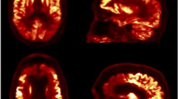
MR-assisted PET data optimization may improve neuroanalysis of dementia patients
Researchers—led by Kevin Chen, PhD, a postdoctoral fellow at Harvard University's Microrobotics Lab—have found that spatiotemporally correlated data acquired using a single magnetic resonance (MR) sequence may be successfully used in a PET/MRI scanner for PET attenuation, motion and partial effects corrections.
The researchers also showed that MR-assisted PET optimization (MaPET) may more accurately assess the pathological changes in patients with dementia and other neurological disorders, according to a study published June 22 in the Journal of Nuclear Medicine.
"A main advantage of PET is that it provides quantitative measures of the radiotracer concentration, but its accuracy is confounded by factors including attenuation, subject motion and limited spatial resolution," Chen et al. wrote. "Using the information from one simultaneously-acquired morphological MR sequence with embedded navigators for MR motion correction (MC), we propose an efficient method, MR-assisted PET data optimization (MaPET), for attenuation correction, PET, MC and anatomy-aided reconstruction."
For attenuation correction, the researchers developed voxel-wise linear attenuation coefficient maps using an SPM8 based method on the MR volume, according to study methods. These embedded navigators were then use for head motion estimates for event-based PET motion correction and region-based analysis were done to assess the quality of the optimized PET images.
Overall, the researchers found the optimized PET images reconstructed with MaPET had better image quality compared to images reconstructed using attenuation correction, even if a patient exhibited large head movements during the imaging scan.
The images acquired were also shown using the Cohen’s d metric to achieve a greater effect size in distinguishing cortical regions with hypometabolism from regions of preserved metabolism, according to the researchers.
"The Cohen’s d metric used to quantitatively evaluate the improvement in image quality suggests that the images are indeed of higher quality after anatomy aided reconstruction (AAR)," the researchers wrote. "As noisy images make the human visual object recognition task more challenging, evaluating the AAR images with reduced noise could potentially improve clinicians’ diagnostic confidence."

