NIH merges microscopes to generate clearer images of processes inside cells
Scientists at the National Institute of Biomedical Imaging and Bioengineering (NIBIB) announced May 7 they had merged two microscope technologies to generate clearer images of rapid processes occurring inside human cells.
Their research was published on May 7 in Nature Methods, according to a NIH press release.
The multifaceted microscope is an improvement to traditional Total Internal Reflection Flourescence (TIRF) microscopy, explained Hari Shroff, PhD, explained chief of NIBIB’s lab section on High Resolution Optical Imaging.
Though TIRF microscopy can create high contrast images by minimizing out-of-focus light, TRIF's ability to improve image resolution takes time and can blur images of small cell features.
In response, Shroff and his colleagues modified a high-speed, super resolution microscope to perform like a TIRF microscope. By combining the two types, the researchers were able to observe processes within cells roughly 10 times faster than other microscopes at similar resolutions.
“TIRF microscopy has been around for more than 30 years and it is so useful that it will likely be around for at least the next 30,” Shroff said in a prepared statement. “Our method improves the spatial resolution of TIRF microscopy without compromising speed—something that no other microscope can do. We hope it helps us clarify high-speed biology that might otherwise be hidden or blurred by other microscopes so that we can better understand how biological processes work.”
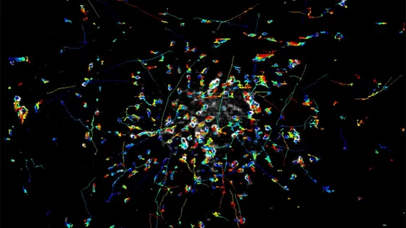
The researchers were able to follow rapidly moving Rab11 particles near the plasma membrane of human cells, according to the release, which otherwise would look blurry. They also could identify the dynamics and spatial distribution of HRas, a protein that responsible for facilitating the growth of malignancy.

