‘Super-resolution’ PET utilizes unwanted movements to bolster brain imaging
A new “super-resolution” molecular imaging technique creates extremely detailed brain scans that may allow for earlier detection of neurological disorders.
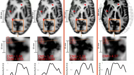
Brain PET imaging quality is typically limited by unwanted movements. But for this investigation, researchers utilized these head motions to enhance exam quality, according to a study presented during the Society of Nuclear Medicine and Molecular Imaging’s annual gathering, which concludes on June 15.
"Our technique not only compensates for the negative effects of head motion on PET image quality, but it also leverages the increased sampling information associated with imaging of moving targets to enhance the effective PET resolution,” said Yanis Chemli, MSc, a PhD candidate at the Gordon Center for Medical Imaging at Massachusetts General Hospital in Boston.
The approach combines PET scanning with a tracking device to capture movement with extraordinarily high precision, the authors noted. After imaging both moving phantoms and primates, the combined data produced scans with “noticeably” higher resolution compared to static reference images.
So far the Boston team has only completed preclinical tests but they’re currently working to include human subjects. For those trials, Chemli and colleagues will likely key in on tau protein tangles in patients with early-stage Alzheimer's.
“The better we can image these small structures in the brain, the earlier we may be able to diagnose and, perhaps in the future, treat Alzheimer's disease," Chemli concluded.

