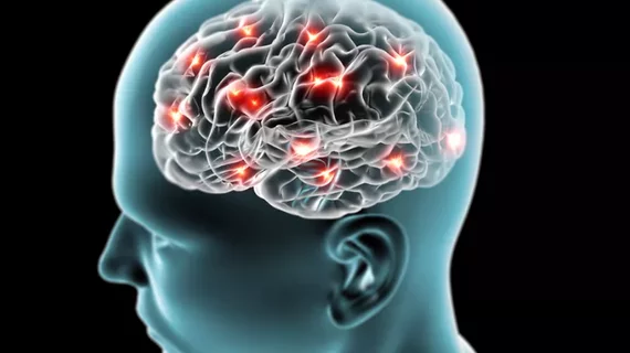
'Queen of Pain' captures neural networks with fMRI
For the average person, pain carries negative connotations—and rightly so—because of intricate connections to fear and anxiety. But one neuroscientist has dedicated her entire life to studying pain, and its neural patterns that can be seen through brain fMRIs.
Irene Tracey, known as the "Queen of Pain," is the director of Oxford University's Nuffield Department of Clinical Neurosciences, PhD, according to an editorial published in the July 2 issue of the New Yorker.
Tracey has designed an imaging-analysis software using fMRI technology to analyze and map with spots of color the neural landscape of pain in several thousand people to date, according to the article.
"Rather than just seeing that all these blobs are active because it hurts, we wanted to understand what bit of the hurt are they underpinning?” Tracey said in an interview with the New Yorker. “Is it the localization? Is it the intensity? Is it the anticipation or the anxiety?”
Most of her research has focused on “good” pain, which comes from an external source and is essential to understanding basic neurobiology. But Tracey has recently shifted her focus to examining "bad" or chronic pain, which affects 10 to 30 percent of Americans.
"Her findings have already changed our understanding of pain; now they promise to transform its diagnosis and treatment, a shift whose effects will be felt in hospitals, courtrooms, and society at large," according to the article.
Read the New Yorker's entire article here:

