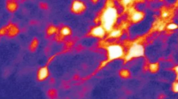
'See-through' EEG allows simultaneous neuroimaging and brain activity monitoring
Although an electroencephalogram (EEG) is a standard method of measuring brain activity, it cannot distinguish differences in types of electrical activity in brain cells. A “see-through” EEG, though, may help provide future insight into neurological conditions such as traumatic brain injury, spinal cord injury and epilepsy.
In the study, Michela Fagiolini, PhD, a neuroscientist and Boston Children’s Hospital and assistant professor of neurology at Harvard Medical School, and Hui Fang, PhD, an assistant professor of electrical and computer engineering at Boston's Northeastern University, captured electrical activity of brain neurons in mice responding to visual stimuli while they simultaneously performing enhanced optical imaging, according to research published online Sept. 5 in Science Advances.
The researchers designed the EEG electrodes to be placed only micrometers apart on the brain cortex, allowing for the collection of information.
Fagiolini explained they also used nanotechnologies that allow light to pass through the EEG microarray, making it transparent. This allows for simultaneous neuroimaging while EEG signals are recorded, according to Fagiolini and colleagues.
The transparent electrodes—engineered into two layers using nanotechnology that are biocompatible and flexible—also allowed the researchers to use genetically modify the cells by using light in conjunction with EEG.
“The highly transparent, bilayer-nanomesh microelectrode arrays allowed in vivo two-photon imaging of single neurons in layer two-thirds of the visual cortex of awake mice, along with high-fidelity, simultaneous electrical recordings of visual-evoked activity, both in the multi-unit activity band and at lower frequencies by measuring the visual-evoked potential in the time domain,” the researchers wrote. “Together, these advances reveal the great potential of transparent arrays from bilayer-nanomesh microelectrodes for a broad range of utility in neuroscience and medical practices.”

