Nuclear Medicine

Nuclear medicine (also called molecular imaging) includes positron emission computed tomography (PET) and single photon emission computed tomography (SPECT) imaging. Nuclear imaging is achieved by injecting small amounts of radioactive material (radiopharmaceuticals) into patients before or during their scan. These can use sugars or chemical traits to bond to specific cells. The radioactive material is taken up by cells that consume the sugars. The radiation emitted from inside the body is detected by photon detectors outside the body. Computers take the data to assemble images of the radiation emissions. Nuclear images may appear fuzzy or ghostly rather than the sharper resolution from MRI and CT.  But, it provides metabolic information at a cellular level, showing if there are defects in the function of the heart, areas of very high metabolic activity associated with cancer cells, or areas of inflammation, data not available from other modalities. These noninvasive imaging exams are used to diagnose cancer, heart disease, Alzheimer’s and Parkinson’s disease, bone disorders and other disorders. 

Video interview with ASNC President President Mouaz Al-Mallah, MD, who explains why nuclear cardiology needs to upgrade its technology to be competitive. #ASNC #ASNC2023 #ASNC23

Previewing ASNC 2023: Why nuclear cardiology needs to evolve

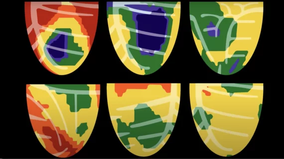
ASNC President President Mouaz Al-Mallah, MD, said nuclear cardiology needs to upgrade old imaging systems and embrace new technology to deliver better value for patients. 

artificial intelligence healthcare industry digest

ChatGPT knows a lot about PET scans, but its advice is inconsistent

The popular AI chatbot got most of the answers it was asked by researchers right, but its reliability is still up in the air.

Thumbnail

Experts advocate for more intensive head-and-neck cancer follow-up using FDG-PET/CT

Scientists believe this is the first series demonstrating a significant survival difference in patients monitored with an intensive follow-up strategy via 18F-fludeoxyglucose–positron emission tomography.  

Tau PET the best tool to predict rate of cognitive decline in Alzheimer’s

Given the approach’s “superior prognostic value,” experts urged for the incorporation of tau PET into routine clinical evaluation in memory care clinics. 

molecular imaging study on brain connections in obese individuals

Obesity linked to altered brain connectivity on PET/MRI exams

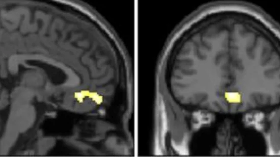
New research, presented at SNMMI 2023, details how the brains of obese individuals differ from those who maintain a healthy weight.  

SNMMI Image of the Year

SNMMI unveils Image of the Year

This year the Henry N. Wagner Jr., Image of the Year is actually a group of images displaying the efficacy of a 68Ga-DOTA-5G and 177Lu-DOTA-ABM-5G theranostic pairing that improves the detection and treatment of metastatic pancreatic cancer. 

whole-body pet scanner add-on device presented at #SNMMI 2023

New PET technology offers 'clear improvement' of whole-body scan resolution

The new technology achieves this by way of two add-on detectors that simultaneously scan patients during their whole-body PET exam.

Thumbnail

New prostate cancer PET imaging agent officially available for commercial use

Posluma (flotufolastat F 18) received the U.S. Food and Drug Administration’s approval in May 2023.

Around the web

GE HealthCare's flurpiridaz, the PET radiotracer that recently received FDA approval, offers several key benefits over SPECT. Jamshid Maddahi, MD, discussed the details in an exclusive interview. 

Ultrafast MCE could go on to become a go-to treatment option for obstructive coronary artery disease, according to the authors of a new first-in-human clinical study.

Elucid's PlaqueIQ was trained to turn CCTA images into interactive 3D reports that help physicians visualize the presence of atherosclerosis.

Trimed Popup
Trimed Popup