Nuclear Medicine

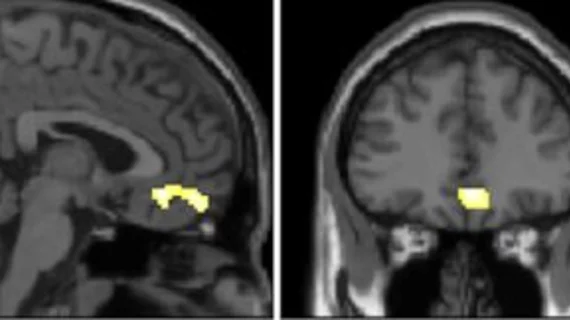
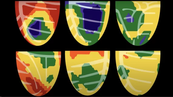
Nuclear medicine (also called molecular imaging) includes positron emission computed tomography (PET) and single photon emission computed tomography (SPECT) imaging. Nuclear imaging is achieved by injecting small amounts of radioactive material (radiopharmaceuticals) into patients before or during their scan. These can use sugars or chemical traits to bond to specific cells. The radioactive material is taken up by cells that consume the sugars. The radiation emitted from inside the body is detected by photon detectors outside the body. Computers take the data to assemble images of the radiation emissions. Nuclear images may appear fuzzy or ghostly rather than the sharper resolution from MRI and CT.  But, it provides metabolic information at a cellular level, showing if there are defects in the function of the heart, areas of very high metabolic activity associated with cancer cells, or areas of inflammation, data not available from other modalities. These noninvasive imaging exams are used to diagnose cancer, heart disease, Alzheimer’s and Parkinson’s disease, bone disorders and other disorders. 

Around the web

Radiologists need better access to patient data stored in the electronic medical record, imaging IT expert Rik Primo explains. 

Imaging IT expert Rik Primo discusses emerging issues he saw at RSNA and HIMSS.

Many new imaging solutions were on display at the world's largest health informatics conference, held at McCormick Place in Chicago.

Cardiologist heart

Advanced imaging does not always improve outcomes, data suggest

Although advanced imaging exams have proven benefits in defining disease severity, new data indicate that more sophisticated studies might not impact outcomes as much as previously thought. 

March 13, 2023

New practice guidelines for PET imaging of neuroendocrine tumors

Issued by the Society of Nuclear Medicine and Molecular Imaging (SNMMI) and the European Association of Nuclear Medicine (EANM), the new guidelines are intended to inform any practitioners involved in ordering, performing, reading and reporting SSTR PET imaging.

February 22, 2023

Amyloid plaque patterns on PET imaging predict Alzheimer's progression in asymptomatic patients

Experts involved in the new research suggest that identifying these spatiotemporal variations could play an important role in clinical research and precision medicine. 

February 8, 2023

E-cigarettes cause worse inflammation than tobacco cigarettes, new PET data shows

The use of e-cigarettes has increased significantly in recent years, mostly due to the fact that many consumers believe them to be a safer option than traditional cigarettes.

January 25, 2023
new radiotracer studies inflammation

New radiotracer could change how radiologists understand inflammation

Labeled as LW223, the fluorinated PET radiotracer is the first of its kind to be impervious to rs6971 polymorphism—a genetic mutation that inhibits the diagnostic view of inflammation imaging.

January 24, 2023

A 'clear lack of adherence' in imaging guidelines is impacting lung cancer survival rates

A study published in the January issue of the Journal of Nuclear Medicine reveals that nearly half of Medicare patients with NSCLC in the United States do not complete recommended FDG PET/CT scans prior to treatment.

January 19, 2023
Blood pressure

‘Revolutionary’ new CT scans identify the most common cause of high blood pressure

Primary aldosteronism (PA) is one the single most common causes of hypertension, but identifying patients with PA—and knowing which ones may benefit from a surgical treatment—can be quite challenging.

January 17, 2023
Blood pressure

New type of PET/CT scan could change how hypertension is diagnosed and treated

The 10-minute scan can “light up” aldosterone-producing nodules in the adrenal glands following an injection of metomidate—a radioactive dye that binds specifically to aldosterone-producing nodules.

January 17, 2023
Trimed Popup
Trimed Popup