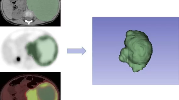
18F-FDG PET/CT radiomics nomogram provides detailed insight into neuroblastoma
A recently developed and validated 18F-FDG PET/CT radiomics nomogram can differentiate between high-risk and non-high-risk neuroblastoma.
Neuroblastoma prognosis varies widely between patients, with some requiring no therapy at all while others develop extensive metastatic disease that deteriorates survival rates. Risk stratification systems, such as the International Neuroblastoma Staging System (INSS), are in place, but are limited in that they most often require surgery or invasive procedures.
For this reason, experts sought to analyze the potential role for radiomics as a means to less invasively identify high-risk patients. Better risk stratification can improve clinical decision making and improve outcomes, experts involved in the study explained.
“Radiomics allows for the assessment not only of shape, including volume, size and area but also of image features, such as patterns and intensity distributions,” corresponding author Jigang Yang, from the Department of Nuclear Medicine at Beijing Friendship Hospital, and colleagues shared. “Radiomics has been widely used for the analysis of pathological lesions and risk stratification in various tumors.”
To develop their nomogram, researchers incorporated clinical factors and a radiomics score (Rad Score). Seven features from 18F-FDG PET/CT images were extracted to construct the signature, which was applied to the imaging of 139 patients (divided into training and validation sets) of the International Neuroblastoma Risk Group (INRG) Staging System (INRGSS).
The age at diagnosis, INRG stage, neuron-specific enolase (NSE) and Rad score, when incorporated all together, yielded an AUC of 0.988 and 0.971 in the training and validation sets for differentiating between high-risk and non-high-risk tumors. Each clinical factor was determined to be an independent predictor of risk.
A decision curves analysis determined the nomogram to be more clinically useful than either the clinical model or the Rad Score alone.
This is the first radiomics nomogram that has been developed to stratify neuroblastoma risk using 18F-FDG PET/CT imaging. Experts involved in the study believe that its use could help patients and clinicians make decisions pertaining to treatment by providing detailed tumor information in a non-invasive manner.
The authors comment:
As a non-invasive quantitative method, it may help the disease follow-up and management in clinical practice and assist in personalized and precise treatment of neuroblastoma.”
The full study can be viewed in the European Journal of Radiology.

