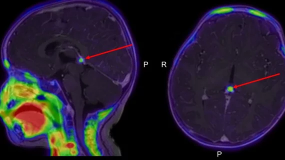
New PET technique slashes scan times, improves image quality in neuroblastoma cases
A new type of PET scan could improve the diagnosis and treatment of neuroblastoma in pediatric patients.
Using a new PET tracer—meta-[18F]fluorobenzylguanidine ([18F]MFBG)—and a long-axial-field-of-view (LAFOV) PET/CT scanner, experts determined that neuroimaging in pediatric patients can be significantly improved. The method reduces scan time and radiation dose, improves image quality and eliminates the need for sedation or anesthesia, authors of a new paper in the Journal of Nuclear Medicine suggest.
“Meta-[18F]fluorobenzylguanidine ([18F]MFBG) has a molecular target similar to that of [123I]MIBG but is radiolabeled with 18F instead of 123I and is therefore different from most of the other PET tracers,” corresponding author Lise Borgwardt, from the Department of Clinical Physiology and Nuclear Medicine at Copenhagen University Hospital in Denmark, and co-authors explain. “It is furthermore a one-day protocol without the need for thyroid-protecting medication.”
Experts recently compared the two techniques in a small group of children who had been diagnosed with neuroblastoma. The new [18F]MFBG PET/CT technique yielded a higher number of radiotracer-avid lesions in 80% of the patients and an equal number of lesions in 20%. What’s more, intraspinal, retroperitoneal lymph node and bone marrow involvement were all diagnosed with much higher precision using the new technique.
None of the children required sedation or general anesthesia, which is typically needed for the standard PET methods used to image neuroblastoma, as they can take two hours or more to complete. In stark contrast, the new technique can be completed in just two minutes without motion.
Combined, the authors suggest that their findings show “promise for future staging and response assessment and may also have a clinical impact on therapeutic decision-making for children with neuroblastoma.”
Learn more about the authors’ findings here.

