Nuclear Medicine

Nuclear medicine (also called molecular imaging) includes positron emission computed tomography (PET) and single photon emission computed tomography (SPECT) imaging. Nuclear imaging is achieved by injecting small amounts of radioactive material (radiopharmaceuticals) into patients before or during their scan. These can use sugars or chemical traits to bond to specific cells. The radioactive material is taken up by cells that consume the sugars. The radiation emitted from inside the body is detected by photon detectors outside the body. Computers take the data to assemble images of the radiation emissions. Nuclear images may appear fuzzy or ghostly rather than the sharper resolution from MRI and CT.  But, it provides metabolic information at a cellular level, showing if there are defects in the function of the heart, areas of very high metabolic activity associated with cancer cells, or areas of inflammation, data not available from other modalities. These noninvasive imaging exams are used to diagnose cancer, heart disease, Alzheimer’s and Parkinson’s disease, bone disorders and other disorders. 

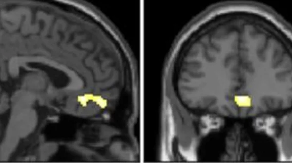
molecular imaging study on brain connections in obese individuals

Obesity linked to altered brain connectivity on PET/MRI exams

New research, presented at SNMMI 2023, details how the brains of obese individuals differ from those who maintain a healthy weight.  

SNMMI Image of the Year

SNMMI unveils Image of the Year

This year the Henry N. Wagner Jr., Image of the Year is actually a group of images displaying the efficacy of a 68Ga-DOTA-5G and 177Lu-DOTA-ABM-5G theranostic pairing that improves the detection and treatment of metastatic pancreatic cancer. 

whole-body pet scanner add-on device presented at #SNMMI 2023

New PET technology offers 'clear improvement' of whole-body scan resolution

The new technology achieves this by way of two add-on detectors that simultaneously scan patients during their whole-body PET exam.

Thumbnail

New prostate cancer PET imaging agent officially available for commercial use

Posluma (flotufolastat F 18) received the U.S. Food and Drug Administration’s approval in May 2023.

folate-based radiopharmaceuticals

Folate-based radiopharmaceuticals could improve detection of gliomas

According to new research, gliomas—a deadly group of brain tumors that are difficult to treat—have increased folate receptor expression, meaning they also show increased uptake of folate-based radiopharmaceuticals on PET imaging.

Thumbnail

Do clinicians want radiologists' management advice? Interviews shed some light on 'unwanted' recommendations

Clinicians only want the information they need to make treatment decisions, rather than advice on what actions they should take, the survey found. 

brain money alzheimer dementia

Will PET imaging be covered alongside new Alzheimer's drugs? CMS dodges topic in new coverage decision

The recent CMS coverage determination did not make any mention of beta-amyloid PET imaging that is necessary for both diagnosing Alzheimer’s and monitoring the effectiveness of related treatments.

Two examples of PSMA-PET scans showing numerous prostate cancer metastases spread throughout the body. Many of these smaller tumors would not have been dected on previous standard-of-care imaging. Photo on left courtesy of SNMMI, right University of Chicago. #PSMAPET

PSMA-PET a more cost-effective option for patients long-term compared to standard prostate imaging

The findings support adopting F-18 DCFPyL PET/CT as the standard of care for prostate cancer staging, authors of a new Scientific Reports paper concluded.

Around the web

Positron, a New York-based nuclear imaging company, will now provide Upbeat Cardiology Solutions with advanced PET/CT systems and services. 

The nuclear imaging isotope shortage of molybdenum-99 may be over now that the sidelined reactor is restarting. ASNC's president says PET and new SPECT technologies helped cardiac imaging labs better weather the storm.

CMS has more than doubled the CCTA payment rate from $175 to $357.13. The move, expected to have a significant impact on the utilization of cardiac CT, received immediate praise from imaging specialists.