3D printed phantom aims to personalize radiation therapy
Meagan Moore, a senior majoring in biological and agriculture engineering at Louisiana State University (LSU) in Baton Rouge has been working for the past year to 3D print a full body “human” phantom for radiation therapy research.
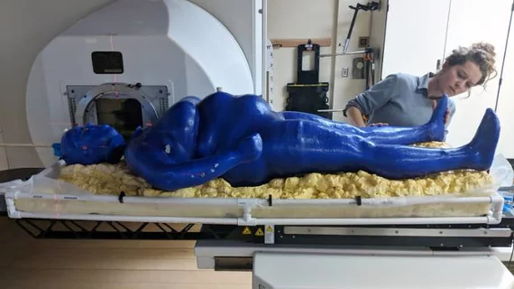
The project, called the Phantom Project or Marie, will test radiation exposure on the personalized, full body phantom to determine the best angle for radiation dose distribution before real-life application and may help personalize cancer treatment for patients of varying body types, according to a recent LSU news release.
Compared to current phantoms which cost thousands of dollars, have no limbs and usually represent one body type, Marie only costs $500, is made of bioplastic that can be filled with water to mimic varying densities of patients and was modeled from 3D imaging scans of five real-life women.
“I specifically wanted to work with a woman because, in science, women typically aren’t studied because they’re considered complex due to a variety of reasons,” Moore said in the release. “I want a person with the most complex geometry.”
Moore spent a total of 136 hours printing Marie into four sections, which she then connected together using soldering, friction stir welding and sandblasting.
Marie’s exterior is a coating of liquid latex and purple roofing sealant for protection. A pipe for dose measurements was placed from the middle of Marie’s head to her pelvic floor. Multiple water tests were conducted to see if 36 gallons of water poured into Marie for four-and-a-half hours could withstand.
The full body phantom has caught the eye of various researchers, including a team from the University of Washington Medical Cyclotron Facility in Seattle who tested fast neuron therapy on Marie.
In the future, Moore hopes replicas of Marie—whose name is a combination of Marie Curie (radiation researcher), Marie Antoinette (symbolized by the detachable head) and Marie Laveau (symbolized by Marie's purple sealant)—will be created and used in the medical field to more precisely treat cancer patients, according to the release.
“What I’d like to see for this project is the research to be used as foundational work to personalize cancer treatments for people with more complex treatments,” Moore said.

