Chest x-rays reveal more severe COVID-19 infections in minority groups
Racial and ethnic minority groups with COVID-19 showed more severe infection on their chest x-rays compared to white/non-Hispanic patients, increasing their chances of dying from the virus, a new study shows.
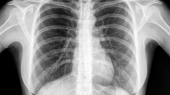
Radiologists from Massachusetts General Hospital analyzed chest radiographs and data from more than 300 patients admitted to their system during the height of the outbreak. The findings—published Thursday in Radiology— align with emerging data that suggests underrepresented racial and ethnic minorities are bearing the brunt of the pandemic, which has now claimed nearly 138,000 American lives.
Co-author Efren J. Flores, MD, a radiologist at MGH, said this research can help create algorithms to identify vulnerable populations, calling on imaging experts to step up to the plate.
“Health equity is every medical specialty’s responsibility, but I believe radiology is uniquely positioned to take a bigger role not only in population health but in public health efforts,” he said in a June 16 statement. “Our ability to provide care in different settings is what allowed us to make the clinical observation that patients coming to this one particular clinic and those being admitted to the hospital were presenting with a higher rate of positive findings that were also more severe. That really offered a window into the disparities that were translating into greater disease severity and worse outcomes.”
In early April, clinicians at an MGH respiratory infection clinic in Chelsea, Massachusetts—a city north of Boston home to primarily Spanish-speaking Hispanic communities—noticed many patients’ chest imaging showed more severe infection than other centers. And they wanted to identify the possible causes.
After looking at the 326 patients, Flores et al. found a number of factors contributed to non-white disease severity including, delaying hospital care, higher prevalence of pre-existing comorbidities, and limited English proficiency.
He noted that as information about the disease evolved, much of it was not available in languages other than English, and said: “that lag in availability of actionable health information for non-English speaking individuals was really critical for many patients trying to navigate a complex medical system with a disease from a virus that is so aggressive.”
Flores reiterated that radiology must do its part to help promote health equity, urging the specialty to collaborate with other medical groups, community stakeholders, and public health initiatives to bolster interventions that increase access to care.
“We did this study not only to gain a better understanding of these emerging disparities, but also to discover how we can use this information to craft a better path towards equity together,” Flores concluded.

