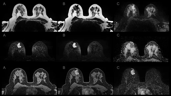
MRI-based radiomics boosts triple-negative breast cancer detection
A new meta-analysis provides detailed insight into the combination of radiomics and breast MRI exams for diagnosing triple negative breast cancer (TNBC).
The paper, published in Clinical Radiology, analyzed six studies comprising 1,223 eligible patients. For each study, researchers calculated pooled sensitivity (SEN), pooled specificity (SPE), positive likelihood ratio (LR+), negative likelihood ratio (LR–), diagnostic odds ratio (DOR), as well as the area under the receiver curve (AUC) to determine the accuracy of MRI-based radiomics for diagnosing TNBC. Their work revealed the method to be an effective diagnostic tool, noting its high specificity for detecting TNBC.
“In routine imaging diagnosis, a lesion can be easily misdiagnosed as benign, thus delaying treatment. Therefore, a non-invasive, early, and accurate diagnostic method for TNBC is urgently needed, and the development of these methods has become a hot topic in the clinic,” explain co-authors Y.S. Sha, of the Department of Radiology at Yantai Hospital of Traditional Chinese Medicine and J.F. Chen, from the Department of Radiology at Southwest Hospital, Third Military Medical University, both in China.
The authors noted that, although MRI has high sensitivity for detecting breast cancer, it is somewhat limited when it comes to identifying TNBC. Prior studies have examined how MRI-based radiomics analyses can be of benefit for TNBC detection, but they lack consistency in size, methodology and model construction, which makes it difficult to validate or discredit and promising results derived from the work.
Due to the aggressive, often invasive nature of TNBC, proper diagnosis and prompt treatment catered specifically to such a diagnosis are pertinent for patients. MRI-based radiomics analysis as a means of predicting molecular status of prostate and brain cancers has been proven to be highly successful, the authors noted. This is what prompted them to dive deeper into the application’s efficacy for diagnosing TNBC.
Upon assessing the six studies that met their inclusion criteria, researchers found that MRI-based radiomics had a pooled sensitivity and specificity of 0.72 and 0.91, respectively. An AUC of .88 was also recorded, indicating that MRI-based radiomics is effective in accurately detecting TNBC, the authors explained.
“This will greatly improve the communication between doctors and patients and facilitate clinical decision-making, and enable the development of optimal treatment plans, and improve clinical outcomes,” the experts said.
They continued by suggesting that the application of MRI-based radiomics for the diagnosis of TNBC could help patients to avoid undergoing invasive, costly biopsies and could also assist providers in obtaining a solid diagnosis more quickly, rather than waiting for histopathologic results.
“Importantly, the present study validated the feasibility of using MRI-based radiomics to identify a disease prone to missed diagnosis and misdiagnosis.”
More on Breast MRI:
Researcher considers greater role of AB-MRI for patients with personal history of breast cancer
Mayo Clinic offers new guidance on supplemental screening of women with dense breasts
Breast MRI screening cuts cancer mortality rates in half for women with lesser-known gene mutations
Abbreviated MRI outperforms DBT for detecting secondary breast cancer

