Tattoo ink can mimic breast cancer on mammography exams
Apparently, some tattoos are more than just skin deep. This was evident in a new case report in Cureus detailing a serious case of mistaken identity on breast imaging.
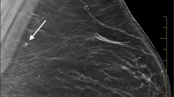
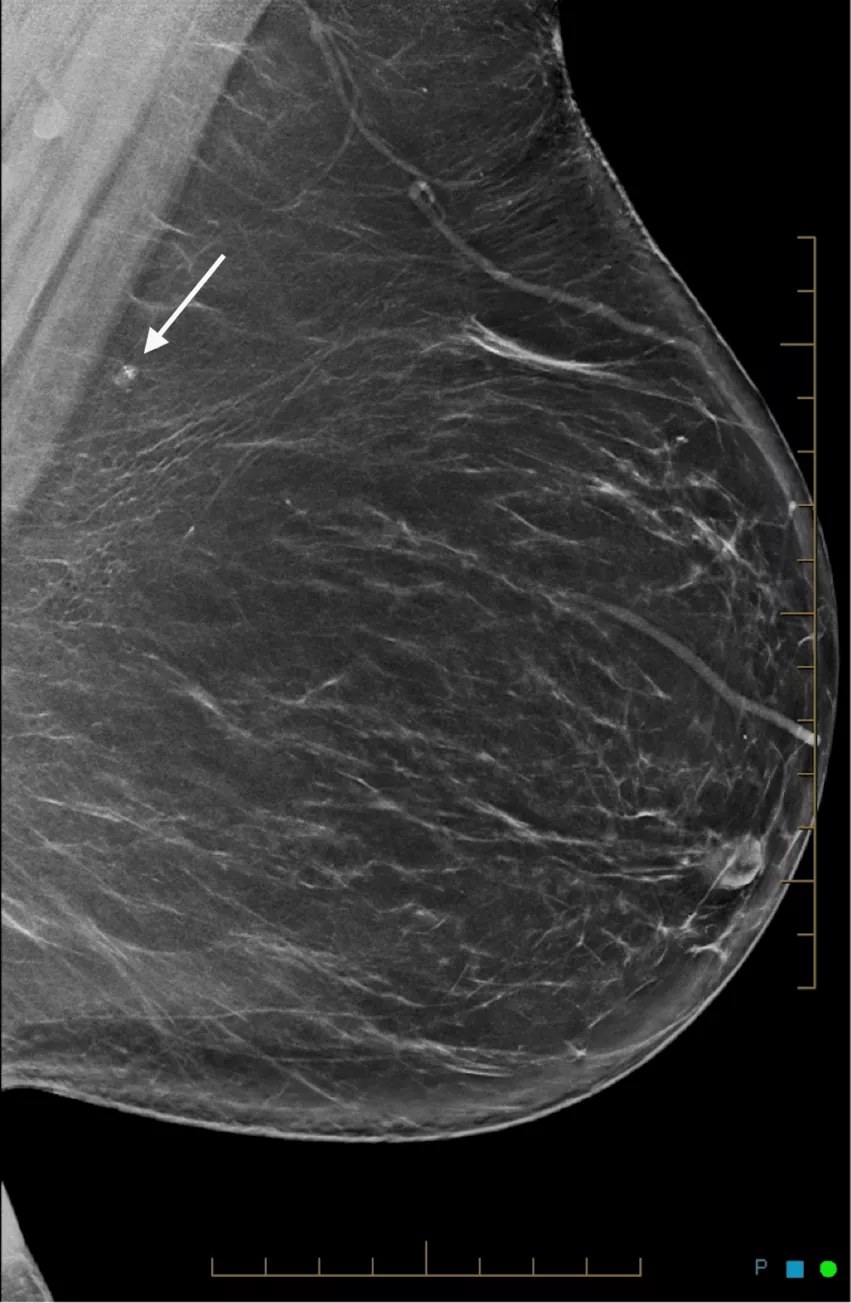
When a 36-year-old BRCA1 mutation carrier female presented for initial screening mammography, radiologists noted a small microcalcification on imaging. The finding was concerning enough to merit further workup, including breast ultrasound and vacuum-assisted biopsy under tomosynthesis guidance, eventually leading experts to a conclusion much less sinister than a cancer diagnosis.
“Mammographic microcalcification has a wide range of etiologies, both malignant and benign. Microcalcification with an intermediate or high probability of malignancy requires further diagnostic workup, such as in the case presented,” corresponding author of the paper Gesine Peters, a medical student at the Tasmanian School of Medicine, and colleagues shared, adding that their findings have not previously been discussed in literature.
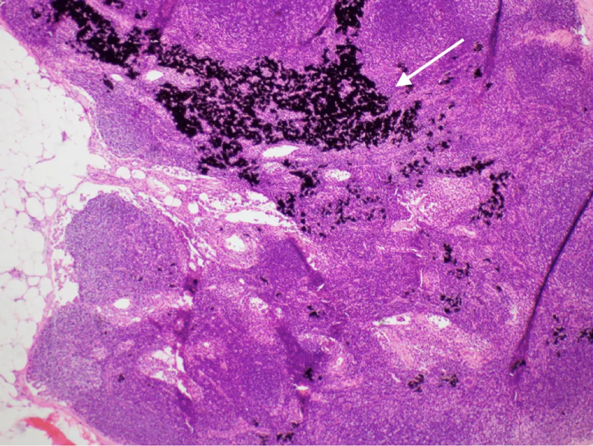
The patient’s breast ultrasound results were normal, leading clinicians to biopsy the area of concern. Those results demonstrated an intramammary lymph node showing prominent extracellular black pigment—revealed to be tattoo ink that had migrated from an upper limb [1].
Different tattoo pigments consist of different materials, including metals, some of which can appear radiopaque on imaging. The authors explained that injuries to the skin during tattoo application can trigger an inflammatory response, which can cause macrophages containing deposited ink to either remain permanently in the skin or be excreted through lymphatic drainage.
Lymphatic drainage of the skin varies by location and individual. The authors shared that while drainage from the trunk to intramammary lymph nodes has been reported on, as has tattoo ink mimicking axillary lymph node calcifications, this is the first time to their knowledge that tattoo pigment has been reported to mimic breast cancer on imaging.
“Uptake of tattoo ink in an intramammary lymph node mimicking a mass lesion containing calcification has not been previously described, however, as the prevalence of females with tattoos increases, tattoo pigment needs to be considered as a potential differential diagnosis of breast calcification,” the authors suggested.
To view more of the images from this case report, click here.


