
How hormonal status affects background parenchymal enhancement on contrasted mammograms
New research details the effects various types of hormones have on breast imaging, highlighting the need for providers to keep radiologists informed on their patients’ use of contraceptives, hormonal therapy treatments, menopausal status and more.
When within a normal range, background parenchymal enhancement (BPE) does not warrant significant concern from radiologists. However, high levels of BPE can indicate an increased risk of breast cancer, especially in women with dense breast tissue.
BPE visualized on breast MRIs is known to be influenced by hormonal status, but less is known about how hormones impact the finding on contrast-enhanced mammography (CEM). A new paper in Radiology is providing detailed insight into how specific factors that impact hormone levels, such as breastfeeding, contraception, medication and menopausal status, alter BPE on CEM.
“Background parenchymal enhancement is an important diagnostic and prognostic imaging biomarker,” Maxine S. Jochelson, MD, with the Department of Radiology at Memorial Sloan Kettering Cancer Center, and colleagues noted. “Although hormonal regulation of BPE at breast MRI has been investigated, information regarding hormonal regulation of BPE at contrast-enhanced mammography remains scarce.”
For the study, researchers retrospectively examined more than 500 CEMs completed between December 2012 and January 2024. The group took note of each patient’s hormonal status, dividing them into five subgroups—premenopausal, postmenopausal, lactating, individuals taking hormone replacement therapy (HRT) and another group treated with tamoxifen. BPE was categorized using the four ordinal grades ranging from minimal to marked.
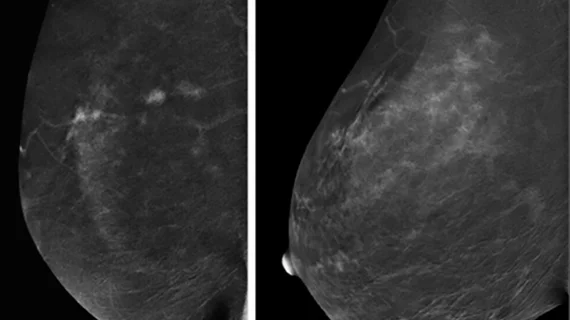
On imaging, premenopausal women displayed higher BPE than those in the postmenopausal group, as did lactating women compared to a group of nonlactating controls; HRT also contributed to higher BPE in comparison to postmenopausal women, while the team observed lower BPE in women treated with tamoxifen compared to a group of nontreated control patients. The group also noted an increase in BPE following the cessation of tamoxifen.
While the authors did not comment on whether the hormonal factors studied affected reader accuracy, they suggested that the “effects should be considered at imaging interpretation” to ensure the reliability of findings.
Learn more about the results here. An accompanying editorial also is available here behind a paywall.

In addition to her background in journalism, Hannah also has patient-facing experience in clinical settings, having spent more than 12 years working as a registered rad tech. She began covering the medical imaging industry for Innovate Healthcare in 2021.