Medical Imaging

Physicians utilize medical imaging to see inside the body to diagnose and treat patients. This includes computed tomography (CT), magnetic resonance imaging (MRI), X-ray, ultrasound, fluoroscopy, angiography,  and the nuclear imaging modalities of PET and SPECT. 

Mercy Medical System first in New York to install clinical LumaGEM MBI system

Gamma Medica announced that New York’s first clinical LumaGEM Molecular Breast Imaging (MBI) system has been installed at Mercy Center, a member of Catholic Health Services of Long Island.

Thumbnail

Gadolinium MRI superior to contrast CT for evaluating some here-and-gone metastases

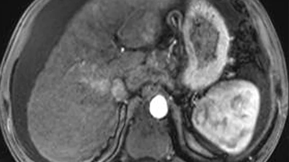
MRI enhanced with gadolinium ethoxybenzyl diethylenetriamine pentaacetic acid—the scan referred to as “EOB MRI”—is significantly better than contrast-enhanced CT for assessing colorectal liver metastases that disappear after chemotherapy, according to a study published online March 22 in Radiology.

Veterinary radiology: The subspecialty no one told you about

The Brookfield Zoo, located just outside of Chicago, uses one of the world’s largest CT scanners to diagnose and treat a wide variety of conditions across the 3,500 animals living at the zoo. The CT scanner was a donation from a local hospital and is capable of imaging animals up to 660 pounds, including fully-grown gorillas, tigers and dolphins.

Thumbnail

Is SPECT imaging accurate in predicting nigral neurons in Parkinson's patients?

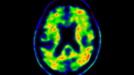
SPECT imaging of the brain is used for gathering information on dopamine activity when it comes to monitoring patients with Parkinson’s disease.

Succeeding with Cancer: Using Imaging to Avoid Treatment-induced Heart Failure

Treating today’s cancer patient no longer means simply targeting the cancer. Given the known cardiotoxicities of some established chemotherapies and the possibility that newer approaches may damage the heart, oncologists, cardiologists and imaging specialists now work together to detect and minimize the risk of treatment-induced heart failure.

Non-invasive device improves cancer monitoring, diagnosis

Diagnosing and monitoring prostate cancer requires patients undergo invasive biopsies. Researchers from Washington State University are changing how physicians do such actions with the development of a new non-invasive, filter-like device. 

Thumbnail

Increased risk of lung cancer, heart attack from radiotherapy for breast cancer patients who smoke

In a new study published in the Journal of Clinical Oncology, researchers found that smokers treated for breast cancer have a higher risk than non-smokers when it comes to developing lung cancer or heart attack as a result of radiotherapy.

Fluid in the lungs being measured by a new technique using ultrasound

Medical researchers and engineers from North Carolina State University have found a new approach that uses ultrasound to measure fluid levels in the lungs.

Around the web

RBMA President Peter Moffatt discusses declining reimbursement rates, recruiting challenges and the role of artificial intelligence in transforming the industry.

Deepak Bhatt, MD, director of the Mount Sinai Fuster Heart Hospital and principal investigator of the TRANSFORM trial, explains an emerging technique for cardiac screening: combining coronary CT angiography with artificial intelligence for plaque analysis to create an approach similar to mammography.

A total of 16 cardiology practices from 12 states settled with the DOJ to resolve allegations they overbilled Medicare for imaging agents used to diagnose cardiovascular disease.