Medical Imaging

Physicians utilize medical imaging to see inside the body to diagnose and treat patients. This includes computed tomography (CT), magnetic resonance imaging (MRI), X-ray, ultrasound, fluoroscopy, angiography,  and the nuclear imaging modalities of PET and SPECT. 

Software biz scores one for mammography by algorithm

A healthcare-software startup in Israel is reporting that it has created a constantly improving algorithm that is better at reading mammography exams “than radiologists using the best computer-aided detection methods.”

Thumbnail

Tempus, University of Michigan develop oncology sequencing program

Health technology company Tempus is partnering with the University of Michigan’s cancer center to bring a new type of personalized treatment for cancer patients to the market.

Thumbnail

Nuclear Science Committee tackles molybdenum supply at September summit

Halloween might be a tad spookier for radiology departments this fall, as the supply of a commonly-used isotope may be restricted in the U.S. when a Canadian reactor ceases production on Oct. 31st. While organizations like the Nuclear Science Advisory Committee have been planning for this closing for years, it held a crucial meeting on the status of molybdenum-99 supply in late September. 

fMRI may bring precision medicine to depression care

Clinical depression is persistently widespread yet notoriously individualized. It often forces patients and therapists to use an arduous trial-and-error approach in search of the right antidepressant at the right dose, if any, along with the right type of talk therapy—again, if any. 

Yes, radiologists are real doctors: 5 reasons why

Oftentimes when someone remarks on whether or not radiologists are “real physicians,” that someone ends up being a physician. Never mind that diagnostic radiologists can rightly claim the mantle of being the doctors’ doctor.

Thumbnail

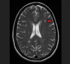
MRI proves worthwhile for assessing MS patients in the emergency room

While it’s not a part of established diagnostic criteria, the use of MRI in the emergency department to evaluate multiple sclerosis (MS) patients for possible exacerbations pays good diagnostic and care-management dividends, according to study conducted at Johns Hopkins and published online Oct. 6 in the American Journal of Neuroradiology.

Thumbnail

New method of gauging metastatic risk may help ovarian-cancer patients avoid unnecessary chest CTs

Researchers in the U.S. and South Korea have collaborated to create and validate an evidence-based rule that can accurately predict which women with ovarian cancer are not at significant risk for metastases in the chest and abdomen—and thus not really in need of some guideline-recommended CT scans.

For dense breasts, 4 screening options after mammograms

Mammograms are the first step when women are on the lookout for breast cancer. But for some women, especially those with dense breasts and who have a special risk according to family history, additional screening might make sense. 

Around the web

RBMA President Peter Moffatt discusses declining reimbursement rates, recruiting challenges and the role of artificial intelligence in transforming the industry.

Deepak Bhatt, MD, director of the Mount Sinai Fuster Heart Hospital and principal investigator of the TRANSFORM trial, explains an emerging technique for cardiac screening: combining coronary CT angiography with artificial intelligence for plaque analysis to create an approach similar to mammography.

A total of 16 cardiology practices from 12 states settled with the DOJ to resolve allegations they overbilled Medicare for imaging agents used to diagnose cardiovascular disease.