Medical Imaging

Physicians utilize medical imaging to see inside the body to diagnose and treat patients. This includes computed tomography (CT), magnetic resonance imaging (MRI), X-ray, ultrasound, fluoroscopy, angiography,  and the nuclear imaging modalities of PET and SPECT. 

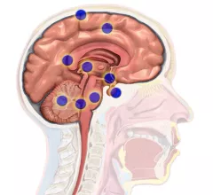
Study pinpoints region of the brain heavily affected by Parkinson's

Researchers have pinpointed regions in the brain that are affected by Parkinson’s disease, opening the door to a potential treatment.

Spot fluoroscopy cuts radiation in image-guided neuro interventions

The innovative imaging technique called “spot fluoroscopy” has shown promise as a way to reduce radiation doses to patients during neurointerventional procedures, as well as scatter doses to staff and clinicians, according to a small study published online Aug. 13 in Acta Radiologica. 

Thumbnail

‘Inevitable’ ioMRI increases brain-tumor procedure times

Intraoperative MRI (ioMRI) soon may be must-have technology for many if not most neurosurgical suites, but those looking to take the plunge for their brain-tumor service line should expect longer procedure times and more scrupulous pre-operative room preparation.

Thumbnail

East Texas Medical Center, Fujifilm continue their long working relationship with transition to DR

Sponsored by FUJIFILM Healthcare Americas

One of the hottest topics in today’s medical imaging community is the transition from computed radiography (CR) to digital radiography (DR).

Thumbnail

Portable DR in the NICU: Safety for the most precious of patients

Sponsored by FUJIFILM Healthcare Americas

Radiation dose is one of the top concerns in imaging today, and since younger patients are the most vulnerable, reducing dose in pediatric imaging is especially important.

Breast cancer imaging challenge one part of achieving Cancer Moonshot

Participants in the Digital Mammography DREAM Challenge are trying to do their part to contribute to the nationwide goal of completing 10 years of cancer research in half the time. It’s funded under the Cancer Moonshot’s Coding4Cancer initiative—pitting coding teams against each other in a friendly fight to see who can come up with the best way to improve mammogram readings. 

Image-based predictor of dementia in patients with Down syndrome

Neural biomarkers could be used to predict the risk for dementia in individuals with Down Syndrome, according to a University of Kentucky study.

Thumbnail

Multiple prior gadolinium doses may remain in numerous brain sections

Gadolinium-based contrast agents (GBCAs) have shown up as bright signals, or “hyperintensities,” in T1-weighted MR brain images of patients with no such contrast administered at present but 35 or more linear GBCA doses in the past. 

Around the web

RBMA President Peter Moffatt discusses declining reimbursement rates, recruiting challenges and the role of artificial intelligence in transforming the industry.

Deepak Bhatt, MD, director of the Mount Sinai Fuster Heart Hospital and principal investigator of the TRANSFORM trial, explains an emerging technique for cardiac screening: combining coronary CT angiography with artificial intelligence for plaque analysis to create an approach similar to mammography.

A total of 16 cardiology practices from 12 states settled with the DOJ to resolve allegations they overbilled Medicare for imaging agents used to diagnose cardiovascular disease.