Medical Imaging

Physicians utilize medical imaging to see inside the body to diagnose and treat patients. This includes computed tomography (CT), magnetic resonance imaging (MRI), X-ray, ultrasound, fluoroscopy, angiography,  and the nuclear imaging modalities of PET and SPECT. 

SPECT can differentiate PTSD from TBI in veterans

Post-traumatic stress disorder (PTSD) and traumatic brain injury (TBI) are common conditions afflicting U.S. military veterans, yet they are difficult to distinguish clinically. A new study using brain perfusion SPECT, however, has demonstrated the ability of the modality to differentiate the conditions.

Risk of cancer related to parathyroid imaging negligible for both 4D CT and scintigraphy

The lifetime risk of developing cancer following parathyroid imaging using either 4D CT or scintigraphy is negligible and should not deter doctors from utilizing the modalities for preoperative assessments, according to study results published in the May issue of the American Journal of Roegentology.

Thumbnail

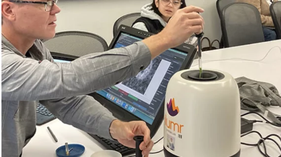
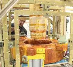
Portable ‘Battlefield MRI’ scanners could bring imaging to war zones, developing countries

The use of MRI has proven to be an invaluable diagnostic tool for assessing injuries in emergency situations—so long as those injured can make it to a hospital, that is. The Battlefield MRI project at Los Alamos Laboratories in New Mexico is aiming to change that.  

Mandatory turnaround times reduce resident involvement in ED imaging

The implementation of turnaround time requirements for imaging reports in emergency departments (EDs) significantly reduces the involvement of radiology residents in the reporting process and may negatively impact their professional training and education, according to study results published in the May issue Academic Radiology.

Thumbnail

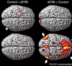
fMRI reveals women suffer more memory impairment following concussion

Women with concussions, or mild traumatic brain injury (MTBI), may have more impairment in working memory processes than men with MTBI, according to a study published online April 28 in Radiology.

Gadolinium retention in brain tissue linked to linear contrast agents

The specific class of gadolinium-based contrast agents (GBCAs) used in prior contrast-enhanced MRI is a determining factor in whether patients exhibit heightened signal intensity on unenhanced T1-weighted MR images, according to study results published online April 6 in the journal Radiology.

Thumbnail

FDG PET not equally valuable for systemic staging of all breast cancer histologies

A recent study has shown that FDG PET/CT, while useful for systemic staging of stage III ductal breast cancer, adds little to the systemic staging of invasive lobular breast cancer (ILC).

Volpara Solutions releases VolparaDensity 3.1 for use with tomosynthesis systems from multiple manufacturers at SBI

Volpara Solutions today announced the release of VolparaDensity version 3.1 here at the Society of Breast Imaging’s SBI/ACR Breast Imaging Symposium in Orlando, Fla., April 25-28, 2015 (SBI Booth #214).

Around the web

A total of 16 cardiology practices from 12 states settled with the DOJ to resolve allegations they overbilled Medicare for imaging agents used to diagnose cardiovascular disease. 

CCTA is being utilized more and more for the diagnosis and management of suspected coronary artery disease. An international group of specialists shared their perspective on this ongoing trend.

The new technology shows early potential to make a significant impact on imaging workflows and patient care.