Medical Imaging

Physicians utilize medical imaging to see inside the body to diagnose and treat patients. This includes computed tomography (CT), magnetic resonance imaging (MRI), X-ray, ultrasound, fluoroscopy, angiography,  and the nuclear imaging modalities of PET and SPECT. 

MRI to 'see through' metal screws developed to follow patients after hip fracture surgery

People who sustain the most common type of hip fracture, known as a femoral neck fracture, are at increased risk of complications. A special type of MRI developed at Hospital for Special Surgery in collaboration with GE Healthcare can show a detailed image following fracture repair, without the distortion caused by metal surgical screws that are problematic in standard MRIs.

CT could predict chemo effectiveness in patients with pancreatic cancer

Routine CT scans that are performed to guide the treatment of pancreatic cancer could predict its effectiveness, according to a study published on Mar. 10 by the Journal of Clinical Investigation. 

Emergency bedside ultrasound exhibits improved sensitivity in urinary stone detection

Emergency department bedside ultrasonography demonstrated better sensitivity as stone size and number increased in a population of patients with CT-proven urinary stones, according to a study published in volume 15 of the Western Journal of Emergency Medicine

Human copper transporter agent effective for both melanoma PET imaging and therapy

A novel theranostic agent has the ability to target melanoma and provide radionuclide therapy in preliminary investigation, according to a study published March 13 in the Journal of Nuclear Medicine.

Varian acquires Velocity clinical oncology data aggregation software

Varian is acquiring assets of Atlanta-headquartered cancer clinic software company, Velocity Medical Solution, including imaging data aggregation software to improve long-term radiation therapy patient management, Varian announced March 12.

Gold marks the spot in new proton beam tumor tracking system

A partnership between Hitachi and Hokkaido University in Tokyo has led to the first molecular tumor-tracking proton therapy system, Hitachi announced today.

Thumbnail

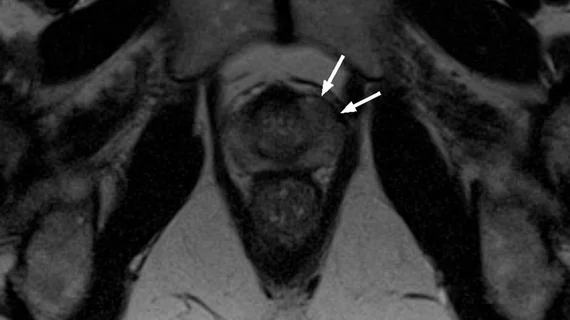
Post-cryoablation MRI features predict liver tumor progression

Researchers have identified MRI features at 24 hours post-cryoablation that were predictive of local liver tumor progression, according to a study published by the American Journal of Roentgenology. These include a minimum ablation margin less than or equal to three millimeters and a blood vessel bridging the margin. 

Alzheimer’s Association grants $8M to Harvard researcher

The largest grant ever provided by the Alzheimer’s Association has been given to Reisa Sperling, MD, a professor of neurology at Harvard Medical School in Boston, to fund its Longitudinal Evaluation of Amyloid Risk and Neurodegeneration study.

Around the web

A total of 16 cardiology practices from 12 states settled with the DOJ to resolve allegations they overbilled Medicare for imaging agents used to diagnose cardiovascular disease. 

CCTA is being utilized more and more for the diagnosis and management of suspected coronary artery disease. An international group of specialists shared their perspective on this ongoing trend.

The new technology shows early potential to make a significant impact on imaging workflows and patient care.