Medical Imaging

Physicians utilize medical imaging to see inside the body to diagnose and treat patients. This includes computed tomography (CT), magnetic resonance imaging (MRI), X-ray, ultrasound, fluoroscopy, angiography,  and the nuclear imaging modalities of PET and SPECT. 

Thumbnail

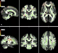
Putative brain iron content indicates changes related to Parkinson’s

Changes related to Parkinson’s disease (PD) were found in putative iron content over two years, according to a study published in the January 2014 issue of Academic Radiology. Rather than correlating with disease duration, the rate of the changes were mildly affected by individual factors like age at disease onset and eventual cognitive decline. 

Thumbnail

DCE-MRI improves accuracy of metastases prediction in breast cancer patients

Integration of DCE-MRI primary lesion kinetics significantly improves the Katz pathologic nomogram accuracy to predict the presence of occult axillary lymph node metastases in breast cancer patients with a positive sentinel lymph node biopsy, according to a study published in the January 2014 issue of Academic Radiology. 

APOE gene variation associated with accelerated brain atrophy

Presence of the epsilon 4 variant of the apolipoprotein E (APOE) gene has been associated with accelerated rates of brain atrophy in individuals with mild cognitive impairment (MCI), according to a study published online Jan. 14 in Radiology.

Rethinking football helmet design

At the University of Florida, a radiologist and mechanical engineer have teamed up to design the next generation of football helmets, created to improve protection from head injuries that are rampant in the sport.

Ketamine boosts serotonin in PET study

The anesthetic ketamine, used in both humans and animals, may have a new use: treating chronic, drug resistant major depression, according to a study published Jan. 7 in Translational Psychiatry.

Isn’t it sweet: FDG may have a rival

The name is fluorodeoxymannose (FDM) and it is giving FDG some competition in the realm of glucose analogue imaging of atherosclerosis, according to a report published Jan. 12 in Nature.

Thumbnail

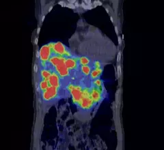
Coming of age for gallium-68 neuroendocrine imaging

Long past are the days when fluorodeoxyglucose was the primary workhorse for imaging neuroendocrine tumors (NETs). Now there is a veritable cornucopia of agents to choose from, but a family of compounds radiolabeled with gallium-68 have been gaining a lot of headway. These are Ga-68 DOTANOC, DOTATOC and DOTATATE.

Future radiation-induced cancer risk negligible in CT of acute TBIs

The lifetime attributable risk of cancer and cancer related mortality is negligibly increased by the radiation emitted from CT use in the evaluation and management of severe traumatic brain injuries (TBIs), according to a study published online Dec. 26, 2013 by the American Journal of Roentgenology. Decisions regarding radiographic evaluation in the acute treatment of TBI should not be influenced by concern regarding future risk of radiation-induced cancer.

Around the web

A total of 16 cardiology practices from 12 states settled with the DOJ to resolve allegations they overbilled Medicare for imaging agents used to diagnose cardiovascular disease. 

CCTA is being utilized more and more for the diagnosis and management of suspected coronary artery disease. An international group of specialists shared their perspective on this ongoing trend.

The new technology shows early potential to make a significant impact on imaging workflows and patient care.