Medical Imaging

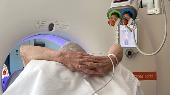
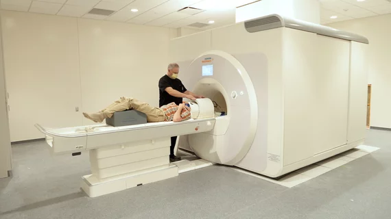
Physicians utilize medical imaging to see inside the body to diagnose and treat patients. This includes computed tomography (CT), magnetic resonance imaging (MRI), X-ray, ultrasound, fluoroscopy, angiography,  and the nuclear imaging modalities of PET and SPECT. 

Genetic test highly sensitive to colon cancer precursors

A noninvasive test to detect cancer precursors in body fluids has been shown to be able to detect genetic variations that initiate colon cancer, potentially signaling an alternative screening method to the standard colonoscopy, according to a study published Sept. 4 in Cancer Prevention Research.

MITA Applauds AATS for Endorsement of Low-Dost CT for High-Risk Lung Cancer Patients

The Medical Imaging & Technology Alliance (MITA) today applauded the American Association for Thoracic Surgery (AATS) for endorsing the United States Preventive Services Task Force’s (USPSTF) draft recommendation for annual low-dose computed tomography scans (LDCT) for individuals at high-risk of developing lung cancer.

Ten outstanding ASTRO members named Fellows

The American Society for Radiation Oncology (ASTRO) has selected 10 distinguished members to receive the Fellow of ASTRO designation (FASTRO). The 2013 class of Fellows will receive the recognition during the Awards Ceremony on Tuesday, September 24, at ASTRO’s 55th Annual Meeting, September 22-25, 2013, at the Georgia World Congress Center in Atlanta.

SNMMI 2013-2015 Wagner-Torizuka Fellowship Recipients Announced

The Society of Nuclear Medicine and Molecular Imaging (SNMMI) is pleased to announce the recipients of the 2013-2015 SNMMI Wagner-Torizuka Fellowship. This two-year fellowship, founded in 2008 by the late Henry N. Wagner, Jr., MD, and the late Kanji Torizuka, MD, PhD, is designed to provide extensive training and experience in the fields of nuclear medicine and molecular imaging for Japanese physicians in the early stages of their careers.

Chasing the lightning: Molecular epilepsy imaging with PET and SPECT

Recent studies have indicated that epilepsy may be more comprehensively characterized with the help of PET and SPECT, and these modalities could benefit the 1-2 percent of the U.S. population with the neurological disorder, according to a review published online Aug 22 in the Journal of Nuclear Medicine.

Thumbnail

Estrogen receptor PET imaging detects early stage breast cancer, with limitations

Early-stage breast cancer lesions can be detected with F-18 FES PET, but success depends on tumor size and level of disease, according to a study published online Aug. 22 in the Journal of Nuclear Medicine.

Optimal contrast dose determined for spotting liver lesions

For the assessment of hypervascular hepatocellular carcinoma (HCC), the optimal iodine dose of contrast material for hepatic arterial-dominant phase (HAP) imaging has been shown to be between 567-647 mgl/kg, according to a study published in the September issue of Academic Radiology.

Radical Radiopharmaceuticals: Molecular Imaging Agents Find Their Niche

Multiple radiopharmaceuticals in the development pipeline are showing real promise in the realms of oncology, neurology and cardiology.

Around the web

RBMA President Peter Moffatt discusses declining reimbursement rates, recruiting challenges and the role of artificial intelligence in transforming the industry.

Deepak Bhatt, MD, director of the Mount Sinai Fuster Heart Hospital and principal investigator of the TRANSFORM trial, explains an emerging technique for cardiac screening: combining coronary CT angiography with artificial intelligence for plaque analysis to create an approach similar to mammography.

A total of 16 cardiology practices from 12 states settled with the DOJ to resolve allegations they overbilled Medicare for imaging agents used to diagnose cardiovascular disease.