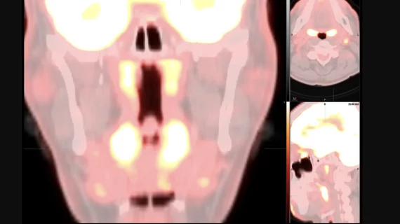
Unusual pattern on PET/CT may indicate COVID omicron variant
A team of nuclear medicine experts has uncovered an “unusual” imaging pattern on radiological scans that may represent omicron. They are now warning providers to be on the lookout when interpreting exams.
The Society of Nuclear Medicine and Molecular Imaging (SNMMI) COVID-19 Task Force put out an update Friday detailing its discovery. Unlike prior COVID strains that primarily manifest in the lungs on PET/CT scans, the new findings center around the upper aerodigestive tract (nose, throat, voice box, etc.) and cervical lymph nodes.
More specifically, radiologists may notice “prominent," symmetric tracer uptake throughout the upper part of the throat behind the nose, middle throat area behind the mouth and tonsils. The pattern may or may not also show associated FDG-avid cervical lymphadenopathy, particularly in the suprahyoid neck, the experts noted.
“The task force recommends that this pattern should be taken into consideration at the time of FDG PET/CT interpretations and the possibility of infection with omicron variant of COVID-19 should be entertained in the differential diagnosis,” the society said in its update.
SNMMI noted that this pattern alone cannot be used to diagnose COVID-19 and offered a handful of suggestions for radiologists to determine if imaging findings do in fact represent infection.
- Look at patients’ records to see if they’ve had a recent positive test result.
- Assess if the individual is more likely to have contracted COVID based on symptoms, recent exposure or travel. If the answer is yes, consider recommending a COVID test.
- Compare the new images with prior PET/CT scans and patient history. The results may represent a chronic inflammatory/reactive process or stable cancer such as lymphoma.
- If the pattern is new, rads should consider many differential diagnoses, including COVID, Epstein-Barr, malignancy and bacterial infection, among other possibilities.
- This new pattern may appear in children and young adults but may simply reflect an increase in normal activity, rather than an underlying illness.
You can read the entire update here.

