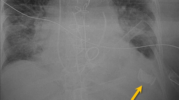
What radiologists should know about spotting items left in patients during surgery
Radiologists must keep an eye out for items left behind in patients during surgery when reviewing chest imaging to prevent deadly complications from arising, according to a new analysis.
The National Quality Forum labels retained surgical items as a preventable complication that should never happen, estimating an incidence of 1.32 cases per 10,000 procedures. These figures, however, are likely higher given organizations don’t often voluntarily report such errors due to legal claims and insurance concerns, experts explained Thursday in RadioGraphics.
RSIs, as they’re known, often appear in the abdomen and pelvis, and show up during thoracic imaging. While rare, they are findings specialists must address quickly.
“Radiologists play a crucial role in early detection of RSIs, which is imperative for timely removal before complications develop,” Gabin Yun, MD, with the University of Michigan Hospital’s Division of Cardiothoracic Radiology, and colleagues explained.
In the postoperative imaging setting, rads should know that a correct sponge or instrument count doesn’t eliminate the possibility that something was left behind. In fact, 88% of incidents occur with correct counts, Yun et al. noted.
Imaging such items may be difficult. Intraoperative radiography can miss 33% of findings, the authors noted, while “radiopaque markers” and a lack of clinical suspicion are also common problems.
Additionally, providers need to be aware of situational risk factors, including an emergency operation, unexpected intraoperative events, longer procedures, high body mass index, multiple operative teams, and poor communication.
The bottom line is organizations and radiology departments must work together to ensure they have a plan in place.
“In addition to using assistive technology for the depiction and counting of RSIs during surgical procedures, various strategies targeting each of the root causes should be implemented organization-wide to reduce the incidence of this preventable complication,” Yun and co-authors concluded.
For more information read the full article here, and watch the entire digital presentation here.

