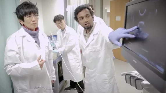
Photoacoustic imaging detects early-stage ovarian cancer
Researchers from Washington University in St. Louis, Missouri used photoacoustic tomography with ultrasound, or photoacoustic imaging, to detect and diagnose early stage ovarian cancer, according to a recent university press release.
Quing Zhu, PhD, professor of biomedical engineering and radiology, at Washington University in St. Louis, and researchers hope the new diagnostic imaging technique could improve early detection in patients with ovarian cancer and help reduce unnecessary surgical intervention, medical costs and improve women's quality-of-life, according to research published online in the September issue of Radiology.
“When ovarian cancer is detected at an early, localized stage—stage 1 or 2—the five-year survival rate after surgery and chemotherapy is 70 to 90 percent, compared with 20 percent or less when it is diagnosed at later stages, 3 or 4,” Zhu said in the statement. “Clearly, early detection is critical, yet due the lack of effective screening tools only 20-25 percent of ovarian cancers are diagnosed early. If detected in later stages, the survival rate is very low.”
Unlike transvaginal ultrasound that tends to lack accuracy, photoacoustic imaging offers information regarding tumor angiogenesis and blood oxygen saturation (sO2) by lighting up the tumor’s vasculature bed. This information, which is related to tumor growth, metabolism and therapeutic response, gives a more accurate diagnosis than conventional methods.
The study involved creating a covering of optical fibers that wrapped around a standard transvaginal ultrasound probe. Once the probe was inserted into the patient, light from the laser spread and was absorbed by the tumor. This ultimately generated sound waves, revealing information about the tumor angiogenesis and sO2 inside the ovaries.
The researchers used two biomarkers to characterize the ovaries, the relative total hemoglobin concentration and mean oxygen saturation. They found the relative total hemoglobin was almost two-fold higher for invasive epithelial cancerous ovaries compared to normal ovaries. The average oxygen saturation of invasive epithelial cancers was nine percent lower than ovaries that are normal or have benign tumors.
“This technology can also be valuable to monitor high-risk patients who have increased risk of ovarian and breast cancers due to their genetic mutations,” Zhu said. “The current standard of care for these women is performing risk reduction surgeries to remove their ovaries at some point, which affects their quality of life and causes other health problems.”
The researchers are planning to apply for funding to conduct a larger clinical trial using the imaging technique, according to the release.

