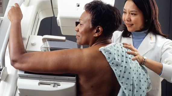
Automated mammogram reveals higher cancer rates in women with dense breasts
A large study utilizing automated mammography revealed a higher cancer rate in women with dense breast tissue compared to those with less-dense breasts.
Previous research reached similar conclusions, but most studies have relied on radiologists’ subjective interpretation using the American College of Radiology’s Breast Imaging Reporting and Data System (BI-RADS). The method leaves an opening for reader variability to influence results.
In this Norwegian study published in Radiology, automated software classified mammographic density in 107,949 women ages 50 to 69 from BreastScreen Norway, a national program offering screening every two years.
Twenty-eight percent of screening exams were classified as dense. Rates of screen-detected cancer were 6.7 per 1,000 examinations in these women, compared to 5.5 for women with non-dense breasts.
Researchers also cited a 3.6 percent recall rate for those with dense breasts compared to 2.7 percent in control. The biopsy rate was slightly higher in the former group (1.4 percent) compared to 1.1 percent in the latter.
The team, led by Solveig Hofvind, PhD, with the Cancer Registry of Norway, also said mammographic sensitivity was reduced in women with dense breasts—reaching up to 71 percent. In those with non-dense breast sensitivity reached 82 percent.
In a Radiological Society of North America (RSNA) release, Hofvind pointed out that more than half of states in the U.S. have passed breast density legislation, but supplemental screening for this group of women is not recommended by any major health care society or organization. She said more research is required before sweeping changes are implemented.
“We need well-planned and high-quality studies that can give evidence about the cost-effectiveness of more frequent screening, other screening tools such as tomosynthesis and/or the use of additional screening tools like MRI and ultrasound for women with dense breasts,” Hofvind said in the release. “Further, we need studies on automated measurement tools for mammographic density to ensure their validity.”

